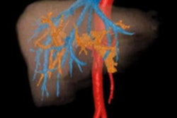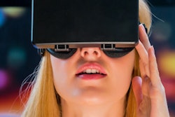
Virtual reality technology could be an effective training tool for interventional radiology (IR), according to a letter to the editor published on 22 July in Academic Radiology.
Why? Because virtual reality-based simulation is a concept interventional radiologists are already familiar with, wrote a team led by Tushar Garg, a medical student at Seth Gordhandas Sunderdas Medical College in Mumbai.
"The technology is better suited for interventional radiology because the image-guided procedures involve viewing video monitors and place the interventional radiologist's hands manipulating instruments at a distance from the actual anatomic site being treated," the group wrote.
Virtual reality systems could help interventional radiologists improve their hand-eye coordination, decision-making, and pretreatment procedure planning, according to Garg and colleagues.
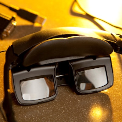
"Immersive environments can be created through panoramic stereoscopic videos, computer-aided design (CAD) modeling, or digital composition via scanning of real environments," they wrote. "[Virtual reality] head-mounted displays can be used to immerse completely in an interventional radiology suite and can be used to [help radiologists] experience basic IR procedures like ultrasonography-guided liver biopsy."
There are obstacles to using virtual reality technology in medical imaging, including its cost, Garg and colleagues conceded. But this particular challenge may resolve on its own.
"The clinical utility of VR in student and patient education as well as in perioperative planning is promising," the authors concluded. "Further technical innovations enabling miniaturization of hardware, decreased costs for adoption and greater reliability of registration of images will allow these technologies to redefine procedural planning and patient engagement."




![A normal mammogram confirmed by three-year radiologic follow-up illustrates reader-marked regions of interest (ROIs) during (A) unaided (round 1) and (B) artificial intelligence (AI)–assisted (round 2) reading. Each colored dot represents an ROI for recall by a human reader. Readers could mark more than one ROI per case, represented by multiple dots of the same color. During AI-assisted reading, the AI system displayed three visible prompts: two with suspicion of malignancy scores of 35% (left mediolateral oblique [L MLO] and craniocaudal [L CC]) and one with a suspicion of malignancy score of 10% (right craniocaudal [R CC]), shown as polygonal overlays. Without AI, six of 10 readers (60%) marked a false-positive ROI. With AI assistance, this fell to two of 10 (20%). R MLO = right mediolateral oblique.](https://img.auntminnieeurope.com/mindful/smg/workspaces/default/uploads/2026/07/2026-07-14-radiology-mammogram-ai-auto-bias.H0bYO8QlWs.jpg?auto=format%2Ccompress&fit=crop&h=100&q=70&w=100)


![A normal mammogram confirmed by three-year radiologic follow-up illustrates reader-marked regions of interest (ROIs) during (A) unaided (round 1) and (B) artificial intelligence (AI)–assisted (round 2) reading. Each colored dot represents an ROI for recall by a human reader. Readers could mark more than one ROI per case, represented by multiple dots of the same color. During AI-assisted reading, the AI system displayed three visible prompts: two with suspicion of malignancy scores of 35% (left mediolateral oblique [L MLO] and craniocaudal [L CC]) and one with a suspicion of malignancy score of 10% (right craniocaudal [R CC]), shown as polygonal overlays. Without AI, six of 10 readers (60%) marked a false-positive ROI. With AI assistance, this fell to two of 10 (20%). R MLO = right mediolateral oblique.](https://img.auntminnieeurope.com/mindful/smg/workspaces/default/uploads/2026/07/2026-07-14-radiology-mammogram-ai-auto-bias.H0bYO8QlWs.jpg?auto=format%2Ccompress&fit=crop&h=112&q=70&w=112)