Dear AuntMinnieEurope Member,
Many radiologists have a love-hate relationship with voice recognition: They know the technology makes their job easier and can boost efficiency, but errors still creep in and every system requires care and attention.
We asked three of our editorial advisory board members to share their experiences and provide some tips on voice recognition. Don't miss our news report. You can find it in the Imaging Informatics Community.
Thoracic CT is an established way of detecting structural pulmonary abnormalities, but it provides no functional information. This prompted researchers at a top U.K. facility to investigate the use of a CT-based full-scale airway network flow model to assess ventilation in chronic obstructive pulmonary disease patients. They compared the modeling results obtained with hyperpolarized xenon-129 MRI and SPECT imaging data. Get the full details.
Contrast-enhanced digital mammography (CEDM) can be used as an alternative modality to contrast-enhanced breast MRI (CE-MRI) in tumor size measurement, a new study published in the British Journal of Radiology has found. What's more, the axial plane of CEDM and CE-MRI should be the first choice for image review and surgical planning. Head over to the Women's Imaging Community.
Meanwhile, Austrian and German investigators have been busy evaluating the impact of magnetic field strength on the accuracy of MRI.
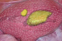
Augmented reality (AR) can provide clinicians with a complete 3D view of subsurface anatomical structures and tumors, and it also allows information from preoperative imaging, such as MRI, to be overlaid and fused in real-time with endoscopic imagery, according to French researchers. They've developed an AR technique that is showing considerable promise. Go to the Advanced Visualization Community.
The European Society for Radiotherapy and Oncology (ESTRO) meeting has taken place in Milan this week. We've posted a news report about a prostate study presented at the event.