Dear Healthcare Informatics Insider,
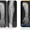
Smartphones are ubiquitous these days, and they are increasingly being utilized to take pictures of patient injuries. These photos can serve as a valuable complement to imaging studies, but they are difficult to incorporate into a patient's record.
Researchers from the University of Applied Sciences of Western Switzerland have developed a prototype mobile app that could make this task a lot easier. Based on HTML5, the Web-based app can securely upload images from any smartphone camera to a database and share them with clinicians.
The photos could also be exported to a patient record if judged to be important, according to the group. Click here to learn more about the prototype.
Speaking of apps, there's been an explosion in the number of imaging-related apps in the past year. But how good are they? It's a mixed bag, according to U.K. researchers. Find out what else they discovered by clicking here.
In other articles in your Healthcare Informatics Digital Community, radiology informatics expert Herman Oosterwijk delves into Clinical Document Architecture (CDA), the document standard defined by HL7 as part of its version 3.
It's a good idea for healthcare imaging and information professionals to get to know CDA, as it's going to be the main "transport" mechanism for clinical information between different systems, according to Mr. Oosterwijk. Find out more in his column here.
Also, a patient radiation tracking project from the International Atomic Energy Agency is making progress and will eventually be operational throughout Europe. Get all the details here.
If you have any tips or suggestions for topics you'd like to see covered in the Healthcare Informatics Digital Community, please feel free to contact me.