Dear Women's Imaging Insider,
A second wave of COVID-19 disease is likely to be coming this autumn or winter, and radiologists must prepare -- particularly those who have close patient contact, such as breast imaging specialists, according to a report published in the European Journal of Radiology Open.
There are a few crucial factors to consider when planning how to continue to provide breast imaging services and keep both staff and patients safe. Find out what researchers from Canada and Singapore suggest in our article posted today.
The regulations concerning the launch of mammography equipment and other devices on the U.K. market are set to change on 1 January 2021, when the Brexit transition period ends. In a timely and practical column, regulatory expert Mike Pogose explains how the new procedures will affect vendors and their customers.
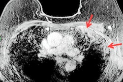
In other women's imaging news, read about a 68-year-old English woman who was found to have COVID-19 after a breast MRI for cancer staging. The woman was asymptomatic for COVID-19 at the time of the MRI, and she only reported feeling mild upper respiratory symptoms two weeks prior to the scan. But the radiologist interpreting her contrast-enhanced MR images found signs of subpleural consolations and a high T2 signal, suggesting she was infected with the novel coronavirus.
Recruitment of women's imaging staff appears to be a particular problem in the U.K. right now. In its annual workforce report, the Royal College of Radiologists has highlighted the skills shortages that are especially acute in certain regions. Don't miss our report on the issue.
These articles make up just part of our Women's Imaging Community reporting. Be sure to check in regularly for current women's imaging news and research developments.




![A normal mammogram confirmed by three-year radiologic follow-up illustrates reader-marked regions of interest (ROIs) during (A) unaided (round 1) and (B) artificial intelligence (AI)–assisted (round 2) reading. Each colored dot represents an ROI for recall by a human reader. Readers could mark more than one ROI per case, represented by multiple dots of the same color. During AI-assisted reading, the AI system displayed three visible prompts: two with suspicion of malignancy scores of 35% (left mediolateral oblique [L MLO] and craniocaudal [L CC]) and one with a suspicion of malignancy score of 10% (right craniocaudal [R CC]), shown as polygonal overlays. Without AI, six of 10 readers (60%) marked a false-positive ROI. With AI assistance, this fell to two of 10 (20%). R MLO = right mediolateral oblique.](https://img.auntminnieeurope.com/mindful/smg/workspaces/default/uploads/2026/07/2026-07-14-radiology-mammogram-ai-auto-bias.H0bYO8QlWs.jpg?auto=format%2Ccompress&fit=crop&h=100&q=70&w=100)


![A normal mammogram confirmed by three-year radiologic follow-up illustrates reader-marked regions of interest (ROIs) during (A) unaided (round 1) and (B) artificial intelligence (AI)–assisted (round 2) reading. Each colored dot represents an ROI for recall by a human reader. Readers could mark more than one ROI per case, represented by multiple dots of the same color. During AI-assisted reading, the AI system displayed three visible prompts: two with suspicion of malignancy scores of 35% (left mediolateral oblique [L MLO] and craniocaudal [L CC]) and one with a suspicion of malignancy score of 10% (right craniocaudal [R CC]), shown as polygonal overlays. Without AI, six of 10 readers (60%) marked a false-positive ROI. With AI assistance, this fell to two of 10 (20%). R MLO = right mediolateral oblique.](https://img.auntminnieeurope.com/mindful/smg/workspaces/default/uploads/2026/07/2026-07-14-radiology-mammogram-ai-auto-bias.H0bYO8QlWs.jpg?auto=format%2Ccompress&fit=crop&h=112&q=70&w=112)