The U.K. Society of Radiographers (SoR) is highlighting research being conducted at Addenbrooke’s Hospital, part of Cambridge University Hospitals, showing that ultrapowerful MRI scans could help patients with epilepsy receive curative surgery.
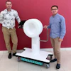
Staff at the Addenbrooke's are currently using a 7 tesla (7T) MRI scanner for research, which is capable of producing images with more detail than standard scanners. They've developed a technique allowing MRI to detect small lesions, noted SoR.
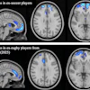
The research described a "triangulation" technique that addresses the issue of signal blackspots on ultra-powerful neuro MR images. Multiple transmitters are positioned around the patient's head in a process known as "parallel transmit MRI." With this, clinicians can detect smaller lesions and allow more patients to be offered surgery to cure their condition.
The study scanned 31 patients with the parallel transmit technique, and 57% of the produced images were clearer than with existing methods, while the rest were of equal quality. As a result, 18 patients are managing their epilepsy differently, including nine who are now able to have potentially curative surgery. The study also found that the procedure was just as comfortable for patients as standard MRI.
The research team is continuing trials using 7T MRI and hopes more patients will benefit from them. For further details, go to: https://www.sor.org/news/mri/ultra-powerful-mri-could-allow-surgery-for-patient


![Overview of the study design. (A) The fully automated deep learning framework was developed to estimate body composition (BC) (defined as subcutaneous adipose tissue [SAT] in liters; visceral adipose tissue [VAT] in liters; skeletal muscle [SM] in liters; SM fat fraction [SMFF] as a percentage; and intramuscular adipose tissue [IMAT] in deciliters) from MRI. The fully automated framework comprised one model (model 1) to quantify different BC measures (SAT, VAT, SM, SMFF, and IMAT) as three-dimensional (3D) measures from whole-body MRI scans. The second model (model 2) was trained to identify standardized anatomic landmarks along the craniocaudal body axis (z coordinate field), which allowed for subdividing the whole-body measures into different subregions typically examined on clinical routine MRI scans (chest, abdomen, and pelvis). (B) BC was quantified from whole-body MRI in over 66,000 individuals from two large population-based cohort studies, the UK Biobank (UKB) (36,317 individuals) and the German National Cohort (NAKO) (30,291 individuals). Bar graphs show age distribution by sex and cohort. BMI = body mass index. (C) After the performance assessment of the fully automated framework, the change in BC measures, distributions, and profiles across age decades were investigated. Age-, sex-, and height-adjusted body composition reference curves were calculated and made publicly available in a web-based z-score calculator (https://circ-ml.github.io).](https://img.auntminnieeurope.com/mindful/smg/workspaces/default/uploads/2026/05/body-comp.XgAjTfPj1W.jpg?auto=format%2Ccompress&fit=crop&h=112&q=70&w=112)


