
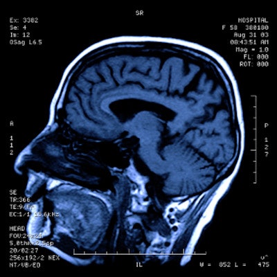
Chronic migraine sufferers show increased brain age on MRI compared with healthy counterparts, according to a study published on 6 October in the Journal of Headache Pain.
The findings could help illuminate the nature of migraine -- a common and disruptive illness, wrote a team led by Dr. Rafael Navarro-González of the Universidad de Valladolid in Spain.
"Migraine is a prevalent and chronic condition known for its recurrent and debilitating headache episodes," the group noted. "Due to the inherent characteristics of migraine and its widespread occurrence, it imposes a substantial burden on both individuals and society as a whole."
Neuroimaging studies have demonstrated that migraine is linked to structural and functional brain alterations, but the association of these changes with aging has not been thoroughly investigated, the team noted. To address this knowledge gap, the group created a machine learning model using the Brain Age Gap Estimation (BrainAGE) framework to evaluate connections between brain age and migraine, hypothesizing that migraine patients would exhibit increased differences between estimated and chronological age compared with healthy peers.
Navarro-González and colleagues trained the model using 2,771 T1-weighted MRI scans of healthy controls. They then used the model with a cohort of 247 patients, of whom 82 were healthy, 91 of whom suffered episodic migraines, and 74 of whom experienced chronic migraines.
The researchers found that chronic migraine patients showed an increased gap between estimated and chronological age compared with healthy controls. The gap for episodic migraine sufferers was also higher compared with healthy controls, but this measure was not statistically significant 1.2 years compared with -0.56 years, p = 0.19).
| Brain age differences (chronological and MRI-assessed) among migraine sufferers compared with healthy controls | |
|---|---|
| Type of migraine |
Brain age gap |
| Chronic migraine sufferers |
4 years |
| Healthy controls |
-0.56 years |
The team did not report any associations between the brain age gap and headache or migraine frequency or duration.
The study indicates that brain-predicted age is a "sensitive biomarker of chronic migraine patients and can help reveal distinct aging patterns in migraine," the group concluded.
"The Brain Age paradigm has shown to be a promising viewpoint for the study of migraine," the group wrote.
The complete study can be found here.


![Overview of the study design. (A) The fully automated deep learning framework was developed to estimate body composition (BC) (defined as subcutaneous adipose tissue [SAT] in liters; visceral adipose tissue [VAT] in liters; skeletal muscle [SM] in liters; SM fat fraction [SMFF] as a percentage; and intramuscular adipose tissue [IMAT] in deciliters) from MRI. The fully automated framework comprised one model (model 1) to quantify different BC measures (SAT, VAT, SM, SMFF, and IMAT) as three-dimensional (3D) measures from whole-body MRI scans. The second model (model 2) was trained to identify standardized anatomic landmarks along the craniocaudal body axis (z coordinate field), which allowed for subdividing the whole-body measures into different subregions typically examined on clinical routine MRI scans (chest, abdomen, and pelvis). (B) BC was quantified from whole-body MRI in over 66,000 individuals from two large population-based cohort studies, the UK Biobank (UKB) (36,317 individuals) and the German National Cohort (NAKO) (30,291 individuals). Bar graphs show age distribution by sex and cohort. BMI = body mass index. (C) After the performance assessment of the fully automated framework, the change in BC measures, distributions, and profiles across age decades were investigated. Age-, sex-, and height-adjusted body composition reference curves were calculated and made publicly available in a web-based z-score calculator (https://circ-ml.github.io).](https://img.auntminnieeurope.com/mindful/smg/workspaces/default/uploads/2026/05/body-comp.XgAjTfPj1W.jpg?auto=format%2Ccompress&fit=crop&h=100&q=70&w=100)


![Overview of the study design. (A) The fully automated deep learning framework was developed to estimate body composition (BC) (defined as subcutaneous adipose tissue [SAT] in liters; visceral adipose tissue [VAT] in liters; skeletal muscle [SM] in liters; SM fat fraction [SMFF] as a percentage; and intramuscular adipose tissue [IMAT] in deciliters) from MRI. The fully automated framework comprised one model (model 1) to quantify different BC measures (SAT, VAT, SM, SMFF, and IMAT) as three-dimensional (3D) measures from whole-body MRI scans. The second model (model 2) was trained to identify standardized anatomic landmarks along the craniocaudal body axis (z coordinate field), which allowed for subdividing the whole-body measures into different subregions typically examined on clinical routine MRI scans (chest, abdomen, and pelvis). (B) BC was quantified from whole-body MRI in over 66,000 individuals from two large population-based cohort studies, the UK Biobank (UKB) (36,317 individuals) and the German National Cohort (NAKO) (30,291 individuals). Bar graphs show age distribution by sex and cohort. BMI = body mass index. (C) After the performance assessment of the fully automated framework, the change in BC measures, distributions, and profiles across age decades were investigated. Age-, sex-, and height-adjusted body composition reference curves were calculated and made publicly available in a web-based z-score calculator (https://circ-ml.github.io).](https://img.auntminnieeurope.com/mindful/smg/workspaces/default/uploads/2026/05/body-comp.XgAjTfPj1W.jpg?auto=format%2Ccompress&fit=crop&h=112&q=70&w=112)


