
Dear MRI Insider,
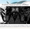
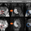
Multiparametric MRI has become a valuable tool for detecting, localizing, and staging of prostate cancer, but the introduction of prebiopsy scans in suspected cases of malignancy has exerted an enormous strain on healthcare systems across Europe. To ensure cancer targets are met, the pressure is on radiology departments to report exams as quickly as possible.
Against this background, radiologists at a teaching hospital in the north of Scotland have embarked on a rigorous campaign to speed up the booking and reporting process. Go to our news report posted today to discover how they've fared.
In other news, an 86-page report issued on 1 October has recommended a large expansion in England's MRI capacity, as well as the training and recruitment of more radiologists and radiographers and the creation of community diagnostic centers.
Among the thousands of journal articles about COVID-19 and radiology published so far in 2020, some papers have focused on the viability of chest MRI as an alternative to chest CT in COVID-19 pneumonia follow-up and on MRI's detection of brain alterations caused by the virus. But has the desire to publish studies quickly resulted in a drop in quality? The Maverinck has expressed his viewpoint in a column.
Meanwhile, German researchers are convinced the stage is set to begin building a strategy for the systematic use of brain MRI scans of breast cancer patients who are at high risk of developing brain cancer metastasis. They presented the results of a new study recently at the European Breast Cancer Conference.
A major challenge in developing artificial intelligence algorithms is having access to a large image dataset with high-quality labels for training. A Swiss team found a way around that problem while developing deep-learning models to detect stroke lesions on MRI.
This letter features only a handful of the many articles posted recently in the MRI Community. Please scroll through the full list below, and feel free to contact me if you have any ideas for coverage.


![Overview of the study design. (A) The fully automated deep learning framework was developed to estimate body composition (BC) (defined as subcutaneous adipose tissue [SAT] in liters; visceral adipose tissue [VAT] in liters; skeletal muscle [SM] in liters; SM fat fraction [SMFF] as a percentage; and intramuscular adipose tissue [IMAT] in deciliters) from MRI. The fully automated framework comprised one model (model 1) to quantify different BC measures (SAT, VAT, SM, SMFF, and IMAT) as three-dimensional (3D) measures from whole-body MRI scans. The second model (model 2) was trained to identify standardized anatomic landmarks along the craniocaudal body axis (z coordinate field), which allowed for subdividing the whole-body measures into different subregions typically examined on clinical routine MRI scans (chest, abdomen, and pelvis). (B) BC was quantified from whole-body MRI in over 66,000 individuals from two large population-based cohort studies, the UK Biobank (UKB) (36,317 individuals) and the German National Cohort (NAKO) (30,291 individuals). Bar graphs show age distribution by sex and cohort. BMI = body mass index. (C) After the performance assessment of the fully automated framework, the change in BC measures, distributions, and profiles across age decades were investigated. Age-, sex-, and height-adjusted body composition reference curves were calculated and made publicly available in a web-based z-score calculator (https://circ-ml.github.io).](https://img.auntminnieeurope.com/mindful/smg/workspaces/default/uploads/2026/05/body-comp.XgAjTfPj1W.jpg?auto=format%2Ccompress&dpr=2&fit=crop&h=167&q=70&w=250)