Dear AuntMinnieEurope Member,
There's been a regular flow of information recently about how alcohol consumption affects the brain. An important new study published today has helped to complete the picture.
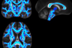
Conventional wisdom is that deterioration of the brain stops when a period of sustained, heavy drinking ends. But using diffusion-tensor MRI, researchers from Spain and Germany have disproved this theory. Go to our MRI Community.
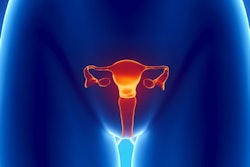
MRI is proving its worth in cervical cancer too. Evaluating parametrial invasion can be essential to determine the most appropriate management of the patient and to improve outcomes, and the most accurate way of doing this is by T2-weighted imaging, according to prize-winning researchers from Italy. They won many plaudits for their work at ECR 2019, and the key findings deserve to be read in the Women's Imaging Community.
Another topic of huge concern for Italian clinicians is how to meet the health needs of the growing number of migrant agricultural workers. Don't miss our report about the efforts of nongovernmental organization Doctors with Africa CUAMM.
Meanwhile, there's good news from the Netherlands. Investigators have used a set of clinical rules to enable many pregnant women suspected of having a pulmonary embolism to avoid unnecessary CT pulmonary angiography scans. Head over to the CT Community.
A research team from a top London facility has developed a convolutional neural network that can determine the make and model of pacemakers and defibrillators within seconds after analyzing radiographs. The deep-learning algorithm proved significantly more accurate and faster than the current flowchart method commonly used by cardiologists to identify such devices.
Last but definitely not least, a month has passed since the end of ECR 2019. Now that you've had time to draw breath, we've produced a short video film about the key trends to take away from the congress in Vienna.


![Overview of the study design. (A) The fully automated deep learning framework was developed to estimate body composition (BC) (defined as subcutaneous adipose tissue [SAT] in liters; visceral adipose tissue [VAT] in liters; skeletal muscle [SM] in liters; SM fat fraction [SMFF] as a percentage; and intramuscular adipose tissue [IMAT] in deciliters) from MRI. The fully automated framework comprised one model (model 1) to quantify different BC measures (SAT, VAT, SM, SMFF, and IMAT) as three-dimensional (3D) measures from whole-body MRI scans. The second model (model 2) was trained to identify standardized anatomic landmarks along the craniocaudal body axis (z coordinate field), which allowed for subdividing the whole-body measures into different subregions typically examined on clinical routine MRI scans (chest, abdomen, and pelvis). (B) BC was quantified from whole-body MRI in over 66,000 individuals from two large population-based cohort studies, the UK Biobank (UKB) (36,317 individuals) and the German National Cohort (NAKO) (30,291 individuals). Bar graphs show age distribution by sex and cohort. BMI = body mass index. (C) After the performance assessment of the fully automated framework, the change in BC measures, distributions, and profiles across age decades were investigated. Age-, sex-, and height-adjusted body composition reference curves were calculated and made publicly available in a web-based z-score calculator (https://circ-ml.github.io).](https://img.auntminnieeurope.com/mindful/smg/workspaces/default/uploads/2026/05/body-comp.XgAjTfPj1W.jpg?auto=format%2Ccompress&fit=crop&h=100&q=70&w=100)


![Overview of the study design. (A) The fully automated deep learning framework was developed to estimate body composition (BC) (defined as subcutaneous adipose tissue [SAT] in liters; visceral adipose tissue [VAT] in liters; skeletal muscle [SM] in liters; SM fat fraction [SMFF] as a percentage; and intramuscular adipose tissue [IMAT] in deciliters) from MRI. The fully automated framework comprised one model (model 1) to quantify different BC measures (SAT, VAT, SM, SMFF, and IMAT) as three-dimensional (3D) measures from whole-body MRI scans. The second model (model 2) was trained to identify standardized anatomic landmarks along the craniocaudal body axis (z coordinate field), which allowed for subdividing the whole-body measures into different subregions typically examined on clinical routine MRI scans (chest, abdomen, and pelvis). (B) BC was quantified from whole-body MRI in over 66,000 individuals from two large population-based cohort studies, the UK Biobank (UKB) (36,317 individuals) and the German National Cohort (NAKO) (30,291 individuals). Bar graphs show age distribution by sex and cohort. BMI = body mass index. (C) After the performance assessment of the fully automated framework, the change in BC measures, distributions, and profiles across age decades were investigated. Age-, sex-, and height-adjusted body composition reference curves were calculated and made publicly available in a web-based z-score calculator (https://circ-ml.github.io).](https://img.auntminnieeurope.com/mindful/smg/workspaces/default/uploads/2026/05/body-comp.XgAjTfPj1W.jpg?auto=format%2Ccompress&fit=crop&h=112&q=70&w=112)


