Dear AuntMinnieEurope Member,
VIENNA - Freezing temperatures and a dusting of snow greeted attendees on the opening day of ECR 2018. It might have been the coldest weather at the congress for more than a decade, but this didn't keep people away. Long registration lines, crowded sessions, and packed public areas were the order of the day.
ECR has grown so much that interventional workshops now take place in the church close to the main U-Bahn exit, and use is also being made of the Sky High Stage on top of the Saturn Tower near the Austria Center Vienna, providing confirmation of the meeting's popularity.
Belgium was the big winner on the opening day of ECR 2018. A group from Leuven was unveiled as the recipient of a magna cum laude award for a new study about radiation dose and body size. Click here for the details.
How radiologists can future-proof themselves with 3D visualization also came under close scrutiny today. Dr. Peter van Ooijen from the Netherlands, Prof. Dr. Thomas Frauenfelder from Switzerland, and others gave some practical advice on this hot topic. To read more, click here.
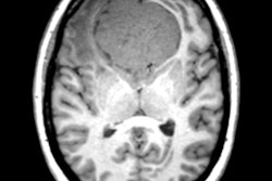
A session on the safety of gadolinium-based contrast agents for MRI also attracted the crowds. Find out more here. And, in a study presented today, French researchers examined the use of gadolinium with MRI scans of patients with intralabyrinthine schwannomas and found that the contrast agent isn't needed for follow-up evaluations. Learn more here.
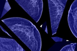
We also have the first two videos from ECR 2018: an exclusive interview with ECR 2018 President Prof. Bernd Hamm about Brexit, artificial intelligence, and attendance numbers and also an interview with Dr. Elisabetta Giannotti on breast imaging and the future of mammography. You won't want to miss them.
Be sure to check our RADCast @ ECR special section throughout the congress to view the live coverage from our five-strong editorial team.


![Overview of the study design. (A) The fully automated deep learning framework was developed to estimate body composition (BC) (defined as subcutaneous adipose tissue [SAT] in liters; visceral adipose tissue [VAT] in liters; skeletal muscle [SM] in liters; SM fat fraction [SMFF] as a percentage; and intramuscular adipose tissue [IMAT] in deciliters) from MRI. The fully automated framework comprised one model (model 1) to quantify different BC measures (SAT, VAT, SM, SMFF, and IMAT) as three-dimensional (3D) measures from whole-body MRI scans. The second model (model 2) was trained to identify standardized anatomic landmarks along the craniocaudal body axis (z coordinate field), which allowed for subdividing the whole-body measures into different subregions typically examined on clinical routine MRI scans (chest, abdomen, and pelvis). (B) BC was quantified from whole-body MRI in over 66,000 individuals from two large population-based cohort studies, the UK Biobank (UKB) (36,317 individuals) and the German National Cohort (NAKO) (30,291 individuals). Bar graphs show age distribution by sex and cohort. BMI = body mass index. (C) After the performance assessment of the fully automated framework, the change in BC measures, distributions, and profiles across age decades were investigated. Age-, sex-, and height-adjusted body composition reference curves were calculated and made publicly available in a web-based z-score calculator (https://circ-ml.github.io).](https://img.auntminnieeurope.com/mindful/smg/workspaces/default/uploads/2026/05/body-comp.XgAjTfPj1W.jpg?auto=format%2Ccompress&fit=crop&h=100&q=70&w=100)


![Overview of the study design. (A) The fully automated deep learning framework was developed to estimate body composition (BC) (defined as subcutaneous adipose tissue [SAT] in liters; visceral adipose tissue [VAT] in liters; skeletal muscle [SM] in liters; SM fat fraction [SMFF] as a percentage; and intramuscular adipose tissue [IMAT] in deciliters) from MRI. The fully automated framework comprised one model (model 1) to quantify different BC measures (SAT, VAT, SM, SMFF, and IMAT) as three-dimensional (3D) measures from whole-body MRI scans. The second model (model 2) was trained to identify standardized anatomic landmarks along the craniocaudal body axis (z coordinate field), which allowed for subdividing the whole-body measures into different subregions typically examined on clinical routine MRI scans (chest, abdomen, and pelvis). (B) BC was quantified from whole-body MRI in over 66,000 individuals from two large population-based cohort studies, the UK Biobank (UKB) (36,317 individuals) and the German National Cohort (NAKO) (30,291 individuals). Bar graphs show age distribution by sex and cohort. BMI = body mass index. (C) After the performance assessment of the fully automated framework, the change in BC measures, distributions, and profiles across age decades were investigated. Age-, sex-, and height-adjusted body composition reference curves were calculated and made publicly available in a web-based z-score calculator (https://circ-ml.github.io).](https://img.auntminnieeurope.com/mindful/smg/workspaces/default/uploads/2026/05/body-comp.XgAjTfPj1W.jpg?auto=format%2Ccompress&fit=crop&h=112&q=70&w=112)


