
Dear AuntMinnieEurope Member,
Every imaging department would love to deliver a high-quality, fast, and efficient service that is also cheap, but this can't be done, says our popular new columnist Dr. Paul McCoubrie. The problem is that quality measures are not quantified in radiology. To read more about this, and his other thoughts on patient contact and communication, click here.
The ECR begins in Vienna next week, and researchers from the Austrian capital are already making the headlines. They have shown that telemammography is more feasible now that low-cost data networks are proliferating and digital mammography equipment is more common. Go to our PACS Digital Community, or click here.
When resources aren't available to implement a CT-based lung cancer screening program, can a scheme using chest radiographs be substituted for at-risk individuals? A multidisciplinary team from northern Italy certainly thinks so. Find out more in our Digital X-Ray Community, or by clicking here.
Kilovoltage (kV) is a key factor in managing radiation dose and contrast in CT, but in clinical practice, kV adaptation can be complex. It's vital to understand kV in standard scan protocols and adapt it in nonstandard individual patients and cases, Dutch researchers believe. Learn more in our CT Digital Community, or click here.
Meanwhile, a group from Jülich, Germany, has made a strong case for multimodal imaging at high field strengths. The team is integrating electroencephalography with a 9.4-tesla MRI/PET hybrid imager, and is getting some promising results. Visit our Molecular Imaging Digital Community, or click here.
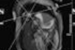
At the recent Society for Cardiovascular Magnetic Resonance meeting, a global group of researchers demonstrated to delegates that cardiac axis localization software can automatically and reliably detect the long-axis views of the heart, offering potential to decrease workload for MR radiographers. Get the story here.


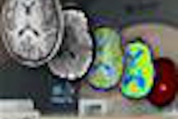


![Overview of the study design. (A) The fully automated deep learning framework was developed to estimate body composition (BC) (defined as subcutaneous adipose tissue [SAT] in liters; visceral adipose tissue [VAT] in liters; skeletal muscle [SM] in liters; SM fat fraction [SMFF] as a percentage; and intramuscular adipose tissue [IMAT] in deciliters) from MRI. The fully automated framework comprised one model (model 1) to quantify different BC measures (SAT, VAT, SM, SMFF, and IMAT) as three-dimensional (3D) measures from whole-body MRI scans. The second model (model 2) was trained to identify standardized anatomic landmarks along the craniocaudal body axis (z coordinate field), which allowed for subdividing the whole-body measures into different subregions typically examined on clinical routine MRI scans (chest, abdomen, and pelvis). (B) BC was quantified from whole-body MRI in over 66,000 individuals from two large population-based cohort studies, the UK Biobank (UKB) (36,317 individuals) and the German National Cohort (NAKO) (30,291 individuals). Bar graphs show age distribution by sex and cohort. BMI = body mass index. (C) After the performance assessment of the fully automated framework, the change in BC measures, distributions, and profiles across age decades were investigated. Age-, sex-, and height-adjusted body composition reference curves were calculated and made publicly available in a web-based z-score calculator (https://circ-ml.github.io).](https://img.auntminnieeurope.com/mindful/smg/workspaces/default/uploads/2026/05/body-comp.XgAjTfPj1W.jpg?auto=format%2Ccompress&dpr=2&fit=crop&h=167&q=70&w=250)