Dear MRI Insider,
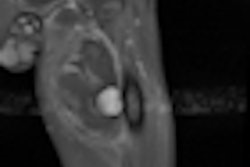
Delegates who made the trek to Bordeaux, France, for last week's International Symposium on State-of-the-Art Imaging (iSi 2011) were treated to a master class in modern applications of neuro MRI by Dr. Birgit Ertl-Wagner. Understanding brain development is essential to diagnosing congenital malformations that often don't become apparent or are missed in childhood, and she urged attendees to seek to improve their anatomical knowledge in order to know where to look for abnormalities.
Our associate editor Frances Rylands-Monk was in the audience, and you can click here to read her report and view some striking images, or visit the MRI Digital Community.
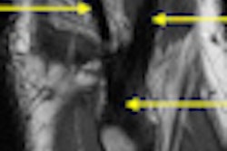
In another iSi 2011 presentation, Dr. Edwin Oei, PhD, discussed how 3-tesla MRI enables doctors to visualize and recognize ligaments in the knee and the smaller joints, such as the posterolateral corner. If not managed correctly, posterolateral corner injuries can cause chronic pain and instability of the knee, he noted. Click here to read more.
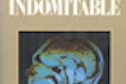
Determining exactly who were the real pioneers in clinical MRI is a topic that has aroused great controversy and emotion during the past three decades or more. The Nobel Committee awarded a medical prize to Paul Lauterbur and Peter Mansfield in 2003, but that was not the end of the saga. Our history column takes a new look at facts. For Dr. Adrian Thomas' latest offering, click here.
Cardiac MRI has not progressed as rapidly as many experts predicted. It's proved tricky to avoid artifacts, but with time and help from physicists and engineers, the technique holds immense promise, Dr. Jeanette Schulz-Menger told delegates at the 2011 Scientific Symposium on Ultrahigh Field Magnetic Resonance in Berlin. For the full story, click here.
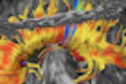
Combining PET with high-field MRI also came under close scrutiny at the Berlin meetting. The technique offers many intriguing possibilities in neuroimaging, including tools to analyze complex neural mechanisms and improve physicians' understanding of neurological disease processes. Our international editor Eric Barnes was onsite, and you can read his report by clicking here.
MRI looks certain to play a central role at the 2012 Olympic Games, but the organizers are eager to sign up extra MR radiographers as volunteers. To find out more, click here. For an in-depth article about imaging of the Olympics, click here.


![Overview of the study design. (A) The fully automated deep learning framework was developed to estimate body composition (BC) (defined as subcutaneous adipose tissue [SAT] in liters; visceral adipose tissue [VAT] in liters; skeletal muscle [SM] in liters; SM fat fraction [SMFF] as a percentage; and intramuscular adipose tissue [IMAT] in deciliters) from MRI. The fully automated framework comprised one model (model 1) to quantify different BC measures (SAT, VAT, SM, SMFF, and IMAT) as three-dimensional (3D) measures from whole-body MRI scans. The second model (model 2) was trained to identify standardized anatomic landmarks along the craniocaudal body axis (z coordinate field), which allowed for subdividing the whole-body measures into different subregions typically examined on clinical routine MRI scans (chest, abdomen, and pelvis). (B) BC was quantified from whole-body MRI in over 66,000 individuals from two large population-based cohort studies, the UK Biobank (UKB) (36,317 individuals) and the German National Cohort (NAKO) (30,291 individuals). Bar graphs show age distribution by sex and cohort. BMI = body mass index. (C) After the performance assessment of the fully automated framework, the change in BC measures, distributions, and profiles across age decades were investigated. Age-, sex-, and height-adjusted body composition reference curves were calculated and made publicly available in a web-based z-score calculator (https://circ-ml.github.io).](https://img.auntminnieeurope.com/mindful/smg/workspaces/default/uploads/2026/05/body-comp.XgAjTfPj1W.jpg?auto=format%2Ccompress&fit=crop&h=100&q=70&w=100)


![Overview of the study design. (A) The fully automated deep learning framework was developed to estimate body composition (BC) (defined as subcutaneous adipose tissue [SAT] in liters; visceral adipose tissue [VAT] in liters; skeletal muscle [SM] in liters; SM fat fraction [SMFF] as a percentage; and intramuscular adipose tissue [IMAT] in deciliters) from MRI. The fully automated framework comprised one model (model 1) to quantify different BC measures (SAT, VAT, SM, SMFF, and IMAT) as three-dimensional (3D) measures from whole-body MRI scans. The second model (model 2) was trained to identify standardized anatomic landmarks along the craniocaudal body axis (z coordinate field), which allowed for subdividing the whole-body measures into different subregions typically examined on clinical routine MRI scans (chest, abdomen, and pelvis). (B) BC was quantified from whole-body MRI in over 66,000 individuals from two large population-based cohort studies, the UK Biobank (UKB) (36,317 individuals) and the German National Cohort (NAKO) (30,291 individuals). Bar graphs show age distribution by sex and cohort. BMI = body mass index. (C) After the performance assessment of the fully automated framework, the change in BC measures, distributions, and profiles across age decades were investigated. Age-, sex-, and height-adjusted body composition reference curves were calculated and made publicly available in a web-based z-score calculator (https://circ-ml.github.io).](https://img.auntminnieeurope.com/mindful/smg/workspaces/default/uploads/2026/05/body-comp.XgAjTfPj1W.jpg?auto=format%2Ccompress&fit=crop&h=112&q=70&w=112)


