Clinical acceptance of two emerging techniques -- irreversible electroporation (IRE) and PET/MRI -- seems to be rising sharply, judged by upbeat presentations given at the 94th German radiology congress, DRK, which ended in Hamburg on Saturday.
Dr. Christian Stroszczynski, a professor of radiology at Regensburg University Hospital presented showed how IRE is showing great promise for the local treatment of cancer. It exposes the tumor to a very brief and intense electric current that leads to the death of the cancer cells, and a key advantage is that the healthy surrounding tissue is spared. To date, there have been no systematic studies of IRE because the procedure is still very new and few centers have clinically tested it, but he is convinced that IRE will become established.
Electroporation is used at present to treat liver tumors, Stroszczynski noted. The liver is frequently affected both by primary tumors (hepatocelluar carcinoma) and metastases, and is well suited to local, minimally invasive therapies such as those used in interventional radiology for some years and with growing success.
"The liver is easily accessible with our working equipment, the probes introduced through the skin (percutaneously), and also the tissue is relatively robust and easily viewed anatomically," he explained.
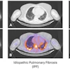
 Above: MRI of an active liver metastasis near the gastric wall (parts of the liver had already been surgically removed two years previously). Below: Three IRE probes surround the tumor. Bottom: Signs of a tissue-sparing destruction of the liver metastasis in the CT review after the procedure.Images courtesy of Dr. Christian Stroszczynski, Regensburg University Hospital.

The interventional radiologist applies two to six probes to the main focus of the cancer. Imaging guidance, typically with CT, is used for navigating the probes, and once the scene of events is reached, a very strong electrical current -- of several thousand volts -- is applied to the tumor tissue, for a fraction of a second. The violent electrical pulse causes the membranes to open and the cells burst.
"This process is equivalent to an induced natural cell death, apoptosis," he said. "Whereas with thermal ablation procedures, for instance, the heat impact also involves the neighboring tissue, there is much less effect on the 'extracellular matrix,' as it is called, which surrounds the cell as a more stable tissue framework, and this usually recovers completely. This especially concerns blood vessels and lymphatic vessels and the nerve pathways. IRE does not induce the tissue necroses, which we have to accept with procedures that involve heat."
Experience so far makes Stroszczynski confident that the procedure will become popular with other centers and that the method can be extended to other organs and cancers, particularly the treatment of prostate cancer, in which it is essential to spare the adjacent tissue. As a general rule, patients can leave the hospital the following day.
PET/MRI of gynecological tumors
Also at DRK 2013, researchers from Essen University Hospital gave a presentation about whole-body PET/MRI with FDG-18 versus whole-body PET/CT in the diagnosis of recurrent gynecological tumors of the pelvis minor, or true pelvis. This showed the value of the method for soft-tissue tumors in particular. Researchers from Essen University Hospital presented the pilot study, which included 12 patients with ovarian and cervical cancer.
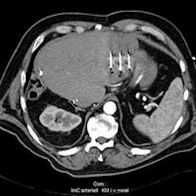
 PET/CT (left) and PET/MR (right) images of patient with metastasising carcinoma of the ovaries. Image courtesy of Dr. Karsten Beiderwellen, Essen University Hospital.
PET/MRI is technically quite demanding, according to Dr. Karsten Beiderwellen, from the Institute of Diagnostic and Interventional Radiology at Essen. For example, it is necessary to ensure that the strong MRI magnetic fields do not interfere with PET diagnostic procedures. Conversely, the PET components must be designed so that their elements are MRI-compatible. He explained that PET/MRI has advantages over the combination of PET and CT.
"First, MRI gives us a better soft-tissue contrast. This is particularly helpful for smaller tumors in soft tissues like the liver, and also in the brain, because the differentiation from the surroundings is much better," he explained. "With PET/CT, we take the CT and the PET one after the other and then have to put them together. With PET/MRI this is done simultaneously. This is a particular advantage with the pelvis minor, because the bladder fills during the investigation and this displaces the adjacent structures somewhat."
Furthermore, the lower radiation burden is a point in favor of PET/MRI.
The Essen team is now working on a direct comparison between PET/MRI and PET/CT in patients with ovarian and cervical cancer. Both organs lie within the pelvis minor, and this region is well suited to PET/MRI because many soft-tissue structures there are closely adjacent. Of 12 patients in the study, all were clinically suspected of having a recurrence of their cancer. Over time, it became apparent that only 10 of them did in fact have a recurrence. The patients were first investigated with PET/CT and directly afterward with PET/MRI.
"So far we have managed to identify all tumor recurrences reliably with both methods. However, small lesions of the liver and also some soft-tissue lesions which were not clear were better characterized with PET/MRI," he noted.
The researchers now eagerly await further results. They want to investigate a total of 50 patients. A special focus of the investigations that will now follow will be the evaluation of tumor secondaries in the lung, an organ which has so far been difficult to access with MRI.