CT alone is not enough to unseat invasive coronary angiography as the gold standard for assessing coronary artery disease, but combining CT with a modality such as SPECT or PET serves as a predictive gatekeeper that can replace unnecessary diagnostic angiography and help avoid ineffective revascularization.
At the recent congress of the European Society of Cardiology (ESC), Dr. Aju Paul Pazhenkottil, a cardiac imaging specialist from Zurich University Hospital, demonstrated the predictive value of SPECT/CT for fusing myocardial perfusion imaging from SPECT with the complementary anatomical CT image.
Pazhenkottil is the lead author of "Prognostic value of cardiac hybrid imaging integrating single-photon emission computed tomography with coronary computed tomography angiography," published in June 2011 in the European Heart Journal (Vol. 32:12, pp. 1465-1471), which was among the first studies to demonstrate the predictive value of combining perfusion and anatomical data.
For the study, 324 patients were divided into three types according to the results of the hybrid exam, a matched group with a finding of stenosis by coronary CT angiography (CTA) and a matching reversible SPECT defect, patients with unmatched coronary CTA and SPECT finding, and then patients found to be normal by both coronary CTA and SPECT.
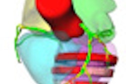
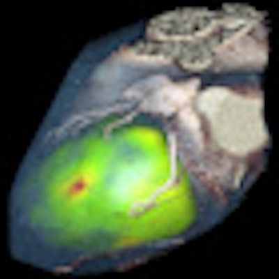
 Hybrid techniques are making steady progress in cardiac imaging. In these cardiac PET/CT images, FDG was used as the radiotracer, along with coronary CTA. Above: Multiplanar reconstruction of fused image. Below: 3D reconstruction. All images courtesy of Dr. Gerald Antoch, department of diagnostic and interventional radiology, University Hospital Duesseldorf, Germany.
At a median follow-up of 2.8 years, a corresponding matched hybrid image finding was associated with a significantly higher incidence of death or myocardial infarction (MI), proving to be an independent predictor for major adverse cardiac events. The annual death/MI rate was 6.0% for patients with matched coronary CTA-SPECT findings, 2.8% for those with unmatched results and 1.3%, among those with normal findings.
Pazhenkottil told AuntMinnieEurope.com his group originally studied matched findings for PET and CT, though the assessment of ischemia by PET is not reimbursed in Switzerland. This preliminary work, "Integrated PET/CT for the assessment of coronary artery disease: a feasibility study," was published in the Journal of Nuclear Medicine in June 2005 (Vol. 46:6, pp. 930-935).
The lack of reimbursement "is too bad because PET is much better, more sensitive and specific," he said, adding that several vendors are combining these two techniques for a faster acquisition in a single scan with lower radiation levels. The combined SPECT/CT test is also available on commercial platforms, he noted.
Responding to concerns from the ESC audience over radiation using hybrid examinations, Pazhenkottil said radiation dose was substantially lowered during the course of the study thanks to the introduction of new CT imaging techniques. The reported levels of the effective radiation dose for stress/rest SPECT and myocardial perfusion imaging (SPECT/MPI) was 10.1 mSv. The estimated radiation dose for the coronary CTA was 17.9 mSv ± 5.8 mSv for 248 patients in which helical scanning was used.
After introducing prospective triggering for coronary CTA, the effective radiation dose for the next 70 patients was systematically recorded at 1.9 mSv ± 0.5 mSv.