Dear AuntMinnieEurope Member,
Subspecialize or be damned: That was the dominant feeling within radiology a few years ago. The prevailing view was that opting for a subspecialty is not a choice but a need for every radiologist. But has this trend gone too far?
In his latest guest column, Dr. Chris Hammond makes a persuasive case for generalists. This is a complex topic requiring careful thought and full consideration, so this article is not a short, easy read. Nevertheless, it's an important issue to address.
Dr. Martin Schranz's turbulent start to the year continues. Following his suspension by University Hospital Kerry, Schranz returned to work on Monday. He was reinstated "on restricted activities," but it remains unclear exactly what that means. This might not be the end of the matter.
Predicting the future is a dangerous business, but those ever-shrewd analysts at Signify Research make a good living out of doing just this. They've taken out their crystal ball and issued their top 10 predictions for the year ahead.
Radiology AI expert Kicky van Leeuwen became a co-owner and consultant at Romion Health in November 2023. During her doctoral studies at Nijmegen, the Netherlands, she led an important study into the effectiveness of commercially available AI algorithms. The group's findings have now been published.
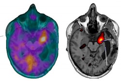
Most epilepsy centers gave up using SPECT seizure imaging some time ago, but the technique may be making a comeback. Swiss researchers have reported that triggering of seizures with direct electric stimulation for ictal SPECT imaging is convenient, spares resources, and can be clinically useful.
Last but definitely not least, many of you will know Dr. Jan Bosmans, PhD, who was an active and valued member of the editorial advisory board of AuntMinnieEurope.com for several years. He died on 13 January, following a long illness. His funeral will take place on Saturday, 20 January at 10 a.m. Central European Time in the Chrysant van crematorium Pontes, Jules Moretuslei 2 in Wilrijk, Antwerp, Belgium. You can follow the farewell via livestream.
Rest in peace, Jan.
Philip Ward
Editor in Chief
AuntMinnieEurope.com


![Examples of ultrasound findings and techniques. (A) Images in a 39-year-old male patient with a mass in the left thigh. The mass is heterogeneous on the B-mode US image (compared with the patient in D) and showed increased microvascularity (superb microvascular imaging [SMI]) and shear-wave elastography (SWE) values. Undifferentiated pleomorphic sarcoma was diagnosed at biopsy (with pleomorphic rhabdomyosarcoma in surgical specimen). (B) Images in an 18-year-old male patient with a mass in the left leg. The mass is hypoechoic on the B-mode image, with no other findings suggestive of malignancy. The lesion is in contact with the cortex of the tibia, which is slightly irregular. CT revealed a doubtful anteromedial tibial erosion. The microvascular study demonstrated high vascularization, suggestive of malignancy. Periosteal Ewing sarcoma was diagnosed with both histologic and immunohistochemical confirmation. (C) Images in a 69-year-old female patient with a lump growing on the outside of the left leg. Multiple SWE examinations were performed (please note the high values obtained in the measurements, whereas the color map highlights the stiffness relative to adjacent tissues). SMI showed areas of increased vascularization to target for sampling. Undifferentiated spindle cell sarcoma was diagnosed at biopsy, with residual leiomyosarcoma in the surgical specimen after neoadjuvant therapy. (D) Images in a 56-year-old female patient with a mass in the right thigh. The mass is heterogeneous at both B-mode ultrasound (similar to patient A) and MRI (coronal T2-weighted spectral attenuated inversion recovery [SPAIR]; T1-weighted pre-contrast and postcontrast imaging), which even shows uptake after the administration of paramagnetic contrast material, which is traditionally suggestive of malignancy. Low values at SMI and elastography are suggestive of benignity. Spindle cell lipoma was diagnosed at biopsy, with atypical spindle cell lipomatous tumor in the surgical specimen.](https://img.auntminnieeurope.com/mindful/smg/workspaces/default/uploads/2026/08/images-radiol250278fig2.APCFLSvX6p.jpg?auto=format%2Ccompress&fit=crop&h=100&q=70&w=100)


![Examples of ultrasound findings and techniques. (A) Images in a 39-year-old male patient with a mass in the left thigh. The mass is heterogeneous on the B-mode US image (compared with the patient in D) and showed increased microvascularity (superb microvascular imaging [SMI]) and shear-wave elastography (SWE) values. Undifferentiated pleomorphic sarcoma was diagnosed at biopsy (with pleomorphic rhabdomyosarcoma in surgical specimen). (B) Images in an 18-year-old male patient with a mass in the left leg. The mass is hypoechoic on the B-mode image, with no other findings suggestive of malignancy. The lesion is in contact with the cortex of the tibia, which is slightly irregular. CT revealed a doubtful anteromedial tibial erosion. The microvascular study demonstrated high vascularization, suggestive of malignancy. Periosteal Ewing sarcoma was diagnosed with both histologic and immunohistochemical confirmation. (C) Images in a 69-year-old female patient with a lump growing on the outside of the left leg. Multiple SWE examinations were performed (please note the high values obtained in the measurements, whereas the color map highlights the stiffness relative to adjacent tissues). SMI showed areas of increased vascularization to target for sampling. Undifferentiated spindle cell sarcoma was diagnosed at biopsy, with residual leiomyosarcoma in the surgical specimen after neoadjuvant therapy. (D) Images in a 56-year-old female patient with a mass in the right thigh. The mass is heterogeneous at both B-mode ultrasound (similar to patient A) and MRI (coronal T2-weighted spectral attenuated inversion recovery [SPAIR]; T1-weighted pre-contrast and postcontrast imaging), which even shows uptake after the administration of paramagnetic contrast material, which is traditionally suggestive of malignancy. Low values at SMI and elastography are suggestive of benignity. Spindle cell lipoma was diagnosed at biopsy, with atypical spindle cell lipomatous tumor in the surgical specimen.](https://img.auntminnieeurope.com/mindful/smg/workspaces/default/uploads/2026/08/images-radiol250278fig2.APCFLSvX6p.jpg?auto=format%2Ccompress&fit=crop&h=112&q=70&w=112)