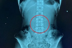
Photon-counting CT (PCCT) can image the lumbar spine at lower radiation dose and without compromising image quality compared with conventional CT, researchers from Zurich have found.
The study results are good news for patient care, wrote a team led by Dr. Adrian Marth of Balgrist University Hospital. The findings were published on 30 August in the American Journal of Roentgenology.
"Lumbar spine CT is recognized to typically have high radiation doses ... [and] the radiation dose of lumbar spine CT may be of particular concern for imaging of pediatric and young adult patients and/or of patients undergoing multiple follow-up examinations," the group explained. "Thus, establishing protocols that provide satisfactory image quality while reducing radiation dose, in accordance with the ALARA [as low as reasonably achievable] principle, will be important as new [PCCT] systems are developed and implemented in clinical practice."
The group conducted a study that included 39 patients who underwent PCCT and 39 patients who underwent a conventional CT exam, both to image the lumbar spine; patients were matched by age, sex, and body mass index.
The exams were performed between June 2022 and January 2023 without contrast but with tin prefiltration (a technique that simultaneously reduces radiation dose and boosts mean photon energy). The researchers then calculated image noise, signal-to-noise ratios, and contrast-to-noise ratios. Three radiologist readers performed visual assessments for each type of exam of study participants' trabecular architecture, cortical bone, neuroforaminal content, paraspinal muscles, and intervertebral disks; they also ranked overall image quality on a four-point scale (1 = poor, 4 = excellent).
| Comparison of conventional CT to PCCT for imaging the lumbar spine | |||
| Measure | Conventional CT | PCCT | p-value |
| Mean CT dose index-volume (CTDI-vol) | 11.1 mGy | 4.4 mGy | p < 0.001 |
| Mean size-specific dose estimate (SSDE) | 14.2 mGy | 6.2 mGy | p < 0.001 |
| Contrast to noise ratio | 29.3 | 33.6 | p < 0.001 |
The group also found that the median score for overall image quality among all three readers was 4 for both types of CT exams.
The findings add to growing evidence that PCCT is an effective addition to the imaging tool kit, the researchers noted.
"The findings support expanded use of [PCCT] for lumbar spine evaluation," they concluded.
The complete study can be found here.