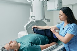
Failure to specify the medical indications in the dose archiving and communication system can increase scan lengths, particularly in cervical and lumbar spine CT exams, a survey of 48 hospitals and clinics in France has revealed.
"The exploration limits proposed for head and abdomen-pelvis examinations by the French guide to radiological procedures (GRP) are consistent with our results. By contrast, for the cervical and lumbar spine, results showed differences between our values and those from the GRP," noted biomedical engineer Luis Alberto González-Méndez, head of studies in medical physics, and colleagues from C2i santé.
"This can be explained by the fact that medical indications were not specified in the dose archiving and communication system (DACS)," added the group from C2i santé, a Nancy-based private medical physics and radiation protection consultancy firm that has worked in CT since 2008.
CT justification
Justifying the length of a CT scan can be complex, depending largely on the clinical indication as well as the experience and awareness of radiographers and radiologists, the authors explained. "This information is not commonly reported in the literature, while few papers have discussed how this parameter can be optimized."
The GRP, introduced by the French Society of Radiology (SFR) in 2006, provides information for professionals on how to perform a CT exam and covers patient preparation, use of contrast media, CT acquisition parameters and patient exposure, etc., but the guide has not been updated since the introduction of the latest generation of CT units, they wrote in a EuroSafe 2023 e-poster presentation.
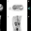
Given this background, they designed a study to determine scan reference length (RL) and guidance length (GL) values for eight of the most common adult CT exams: head, sinus, pulmonary embolism, chest, chest-abdomen-pelvis, abdomen-pelvis, and cervical and lumbar spine.
Data were collected by seven medical physicists between February 2018 and June 2022 in 48 clinics employing a DACS. Most of the DACS exports were performed using Dosewatch (GE Healthcare) and Radiation Dose Monitor (RDM, Medsquare). Data were included and processed in the C2i santé database, containing information on hospital name, public or private facility, anatomical site of CT exam, patient demographics (age, gender, height, weight), CT equipment (model, commissioning year, slice number), acquisition parameters (rotation time, pitch, beam collimation), and exposure data (CTDIvol, DLP).
The retrospective analysis involved 9,180 patients and 55 CT scanners from three large CT vendors (81% of systems came from GE Healthcare, 15% Siemens Healthineers, 4% Canon). The CT exams selected for this study comprised abdomen/pelvis (21%), chest (16%), chest/abdomen/pelvis (16%), head (15%), lumbar spine (11%), pulmonary embolism (10%), sinuses (6%), and cervical spine (5%).
| Imaged scan length results in the head region (cm) | ||
| CT exam | Head | Sinus |
| 75th percentile | 17.5 | 13.5 |
| Maximum | 27.9 | 21.1 |
| Median | 16.5 | 12.4 |
| Minimum | 9 | 7.8 |
| 25th percentile | 15.7 | 11.3 |
| GRPmax | 18 | - |
| GRPmin | 15 | - |
The table above shows that the interquartile explored lengths (25th-75th percentiles) for the head CT examination (16.5 cm) are within the min-max range proposed by the GRP. The sinuses are not included in the GRP.
However, the median imaged scan length for pulmonary embolism exceeds the GRP recommendations by 2.6 cm. The difference in terms of exploration between the PE and the chest examination is 1.5 cm, whereas the GRP recommends the same value (30 cm).
| Imaged scan length results in the trunk region (cm) | ||||||
| CT exam | Pulmonary embolism | Chest | Chest/abdomen/pelvis | Abdomen/pelvis | Cervical spine | Lumbar spine |
| 75th percentile | 35.3 | 36.2 | 67 | 47.5 | 23.1 | 28 |
| Maximum | 47.4 | 48.8 | 92.7 | 54.8 | 49.1 | 40 |
| Median | 32.6 | 34.1 | 64.3 | 44.9 | 20.5 | 25.7 |
| Minimum | 18.2 | 16.3 | 43.5 | 18.3 | 13.8 | 12 |
| 25th percentile | 30.1 | 32 | 61.3 | 41.5 | 18.5 | 23.3 |
| GRP | 30 | 30 | - | - | 15 | - |
| GRPmax | - | - | 80 | 50 | - | 20 |
| GRPmin | - | - | 70 | 40 | - | 15 |
The median imaged scan length for the chest exceeds the GRP recommendations by more than 4 cm. One possible explanation is that these explorations are performed for oncological purposes extending thereby to the adrenal glands and part of the liver to detect possible metastases.
For the cervical spine, the median explored scan length (20.5 cm) exceeds the recommended value in the GRP (15 cm) by more than 5 cm. The maximum value (49.1 cm) is probably a cervicothoracic examination, which was not identified in the DACS. For the lumbar spine, most of the explored scan lengths (25th-75th percentiles) exceed the min-max range values recommended by the GRP of approximately 8 cm. For some patients, the exploration probably involved the dorsolumbar spine and not only the lumbar spine, without being reassigned in the DACS.
"Cervicothoracic or dorsolumbar spine examinations are two examples of existing examinations without specific protocols, therefore spine examination requires an accurate identification," the researchers noted. "The number of vertebrae to be examined will influence the imaged scan length and consequently the DLP. Additionally, lumbar spine examination requires specific identification for diagnostic purposes or examination followed by surgery (arthrodesis)."
| Guide length (GL) and reference length (RL) values per CT examination proposed in the study | ||
| Examination | RL (cm) | GL (cm) |
| Head | 17.5 | 16.5 |
| Sinus | 13.5 | 12.4 |
| Pulmonary embolism | 35.3 | 32.6 |
| Chest | 36.2 | 34.1 |
| Chest/abdomen/pelvis | 67 | 64.3 |
| Abdomen/pelvis | 47.5 | 44.9 |
| Cervical spine | 23.1 | 20.5 |
| Lumbar spine | 28 | 25.7 |
The table above summarizes the imaged scan length values proposed in the study. An RL represents the professional practices of the facilities included in the survey, and it also corresponds to the value not to be exceeded from a regulatory point of view. The GL represents the target to be reached in terms of imaged scan length.
Limitations and the future
The study has limitations, the authors wrote. "Most importantly, medical indications were not specified in the data sample collected. However, this study allowed us to propose eight imaged scan length values, notably a new reference value for the sinus examination, not included in the GRP and the literature."
Future prospective studies are needed to correlate the imaged scan length with patient height and Body Mass Index, they concluded. "In the near future (probably next year) we will publish scan length values for 15 CT exams," González-Méndez told AuntMinnieEurope.com.
To view the entire poster, go to the EPOS section of the ESR website. The co-authors were Isabelle Rousselle (medical physicist), Lisa Palduplin (radiation protection engineer/radiographer, Brice Royer (medical physicist), Alain Noel (medical physicist), and Jad Farah (medical physicist).