A European research group is calling for the use of amyloid PET scans for Alzheimer's disease early in the diagnostic workup of memory clinic patients, according to a study published on 8 May in JAMA Neurology.
A team led by Daniele Altomare, PhD, of the University of Geneva in Switzerland, assessed the clinical effect of amyloid PET in a clinical trial in patients in eight memory clinics. Early PET scans led to more than three times as many patients being diagnosed, they found.
"Widespread implementation of this imaging technique may improve the timely diagnostic workup of patients under evaluation for cognitive decline," the group wrote.
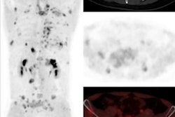
Amyloid PET scans allow for the direct assessment of amyloid deposits in the brain, one of the main hallmarks of Alzheimer's disease. Despite increasing use of the scans in clinical practice, real-world evidence is still limited, in many cases due to the lack of a control group in studies, the authors noted.
To provide evidence to fill this gap, the researchers recruited 844 participants across eight European memory clinics between April 2018 and October 2020. Enrolled patients showed either subjective cognitive decline (plus clinical features increasing the likelihood of preclinical Alzheimer's disease), mild cognitive impairment, or dementia.
The investigators split participants into three groups based on when they underwent amyloid PET: group 1, early in the diagnostic workup (within 1 month); group 2, late in the diagnostic workup (after a mean 8 months); and group 3, if and when the managing physician chose. The main outcome was the difference between group 1 and group 2 in the proportion of participants receiving an Alzheimer's disease diagnosis with very high confidence.
After three months, 109 of 272 participants (40%) in group 1 had a diagnosis with very high confidence versus 30 of 260 (11%) in group 2 (p < 0.001), according to the findings.
"Early amyloid PET allowed memory clinic patients to receive an etiological diagnosis with very high confidence after only three months compared with patients who had not undergone amyloid PET," the group wrote.
The study is one of several to come from the Amyloid Imaging to Prevent Alzheimer's Disease Diagnostic and Patient Management Study (AMYPAD-DPMS), which the group launched in 2018, and is the largest European effort to assess the clinical effect of amyloid PET in memory clinic patients, the researchers noted.
"These findings support the implementation of amyloid PET early in the diagnostic workup of memory clinic patients," the authors concluded.