SPECT cameras with cadmium zinc telluride (CZT) detectors perform comparably to conventional gamma devices for imaging patients with suspected heart attack, Swedish researchers have reported.
The findings cast doubt on the potential advantages of SPECT for this indication, noted a group led by Dr. Fredrik Hedeer, PhD, of Lund University in Lund, Sweden. The study was published on 13 March in the Journal of Nuclear Cardiology.
"Differences between a CZT and a conventional gamma camera for detection of myocardial infarction and assessment of LV [left ventricular] volumes and LVEF [ejection fraction] are small and do not appear to be clinically significant," the researchers wrote.
SPECT myocardial perfusion imaging is widely used to diagnose coronary artery disease by measuring blood flow deficits, yet its ability to detect small infarctions in the endocardium is limited due to image resolution, especially when compared to, for example, standard late gadolinium enhancement MRI, according to the authors.
Recently, however, a new generation of gamma camera systems has evolved that use a solid-state detector technique with more sensitive cadmium-zinc-telluride (CZT) detectors rather than standard sodium iodide (NaI) crystal detectors. With improved spatial and energy resolution, these new cameras may improve the diagnostic performance of MPI, but the comparative performance of the machines for detecting heart attack in patients has not been investigated, the team explained.
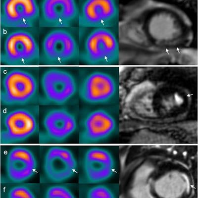
 Patient examples of myocardial infarct (MI) by cardiac MRI, myocardial perfusion SPECT with a cadmium-zinc-telluride (CZT) gamma camera and a conventional (Conv) gamma camera. Columns from left to right show summed myocardial perfusion SPECT images, gated myocardial perfusion SPECT in end-diastole (ED) and end-systole (ES) and late gadolinium enhancement (LGE) cardiac MRI. Case a and b are examples of an infarct in the apical inferior wall on cardiac MRI, which is correctly diagnosed by both CZT and conventional SPECT (arrows). Case c and d are examples of an infarct in the apical lateral wall on cardiac MRI (arrow), missed by both CZT and conventional SPECT. Case e and f are examples of an infarct in the basal lateral wall on cardiac MRI correctly diagnosed by the CZT camera (arrows) but missed by conventional SPECT. Image courtesy of the Journal of Nuclear Medicine through CC BY 4.0.
To address the knowledge gap, the researchers enrolled 73 patients with known or suspected chronic coronary syndrome. Patients underwent both a CZT gamma camera and a conventional gamma camera exam, as well as a late gadolinium enhancement MRI (reference standard). The main outcome was how well the approaches measured left ventricular ejection fraction, which indicates how much oxygen-rich blood is being pumped out of the heart's left ventricle.
Of the patient cohort, 42 were diagnosed with myocardial infarction based on late gadolinium enhancement MRI. The investigators found that performance measures for the CZT and conventional gamma camera were mostly the same:
- Overall sensitivity, 67%
- Specificity, 100%
- Positive predictive value, 100%
- Negative predictive value, 69%
However, for small myocardial infarcts, the CZT system outperformed the conventional gamma device, with a sensitivity of 82% compared with 73%.
The authors did note that the study had limitations, but they emphasized that the research demonstrated a small beneficial edge for the CZT gamma camera, which showed good diagnostic accuracy for detecting myocardial infarction.
"Absolute values of LV volumes and mass should be handled with care in a clinical report of an individual patient, and rather be interpreted compared to specific reference values for each setting, where type of gamma camera, image reconstruction parameters and myocardial perfusion imaging software have been taken into account," the authors concluded.