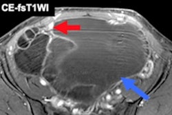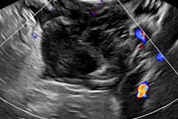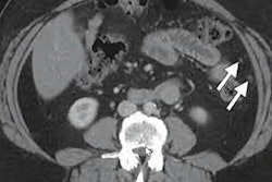
The combination of physical examination, transvaginal ultrasound (TVUS), and pelvic MRI is the best way to diagnose deep infiltrating endometriosis in certain locations, a research team from Paris has reported.
Researchers led by Dr. Alexis Roditis from Pierre and Marie Curie University noted that the combined approach yielded higher sensitivity and accuracy for central locations of endometriosis, such as the vagina, bladder, and uterosacral ligaments.
"Our study demonstrates that a model with at least two concordant positive tests among physical examination, TVUS, and MRI is the most valid approach to diagnose any central location of deep infiltrating endometriosis to maintain the high sensitivity of MRI and reach the high specificity of the physical exam and TVUS," Roditis and colleagues wrote in Fertility and Sterility.
Physical exams have been considered accurate in assessing endometriosis diagnosis by correlating pain with induration or nodules found on digital exams. However, this method has a lower sensitivity when compared with medical imaging techniques such as ultrasound or MRI.
TVUS and MRI are also used in clinical practice. Although MRI is highly sensitive, it is generally less accessible than TVUS. Meanwhile, TVUS has variable sensitivity based on endometriosis location.
Roditis and colleagues wanted to explore the accuracy of the three methods alone and in combination. They looked at retrospective data collected between 2016 and 2020 from 178 women. The women underwent physical exams, TVUS, and pelvic MRI for deep infiltrating endometriosis up to 12 months prior to surgery.
The team tested two model types -- model A and model B -- when combining methods and modalities. Model A refers to all three techniques being positive and concordant, while model B refers to at least two techniques being positive and concordant. All other combinations were considered negative.
The researchers tested these combinations on endometriosis locations, including uterosacral ligaments, vaginal, lateral, and rectosigmoid. They found that a combination model using model B showed the highest sensitivity out of all combinations.
| Diagnostic performance for combinations of physical exam, TVUS, and MRI for deep infiltrating endometriosis | |||||
| Physical exam + MRI | Physical exam + TVUS | TVUS + MRI | Model A | Model B | |
| Sensitivity | 79.4% | 80% | 87.4% | 77.7% | 91.4% |
| Specificity | 33.3% | 33.3% | 66.7% | 33.3% | 33.3% |
| Accuracy | 78.7% | 79.2% | 33.3% | 77% | 90.4% |
For individual methods, MRI showed the highest sensitivity and accuracy at 94.9% and 93.8%, respectively.
MRI showed more sensitivity than physical exam, TVUS, or any combination to detect deep infiltrating endometriosis. The researchers also found that MRI and model B were the most accurate for detecting uterosacral ligaments and rectosigmoid locations. This included accuracies of 90.4% and 82.6%, sensitivities of 91.1% and 50%, and specificities of 77.8% and 90.9%, respectively.
Model B also had the highest accuracy for the vaginal endometriosis location at 82.6%. This also included a sensitivity of 50% and a specificity of 90.9%.
Finally, MRI showed more accuracy than any combination for identifying a lateral location at 75.1%. It also had a sensitivity of 36% and a specificity of 93.8%.
The team concluded that the combined approach (model B) is best for diagnosing any central location of deep infiltrating endometriosis, including uterosacral ligaments, vaginal, rectosigmoid, and bladder. MRI alone meanwhile is best for lateral locations, such as parametrial and sacrorectogenital septa.
Despite their results, the study authors noted that access to all three diagnostic methods is "frequently" limited. They wrote that this is because not all centers are equipped with MRI and the waiting time for an examination can be lengthy. Also, not all radiologists are trained to diagnose endometriosis.
"This point deserves to be underlined because it restricts the application of a combination of physical examination, TVUS, and MRI for the diagnosis of deep infiltrating endometriosis," they wrote in the paper published on 19 December.
The team also called for their results to be confirmed in a screening population.