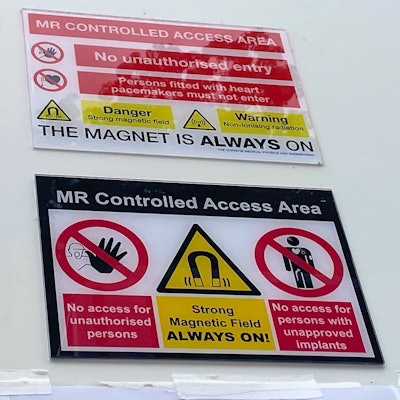
A successful approach to MRI safety must involve a multidisciplinary team effort to arrive at good local decisions, according to Geoff Charles-Edwards, PhD, head of MR physics at Guy's & St Thomas' NHS Foundation Trust in London.
"MR safety is not binary," he told delegates at the British Institute of Radiology (BIR) annual congress. "There is always some sort of risk associated with pretty much everything, so the appropriate way really is to try and understand what the risk is, weigh it up against the perceived benefits, and try and manage it in that situation. To make that call you need to understand both the risks and clinical need -- that itself is quite a challenge."
Risk assessments should be overseen by a safety expert/technician who obtains input from radiologists, radiographers, referring clinicians, and other specialists, said Charles-Edwards, who is a consultant clinical scientist at Guy's & St Thomas' and an honorary senior research fellow at King's College London.
Clear and visible warning signs have an essential role to play in MRI safety.Cardiovascular devices and MRI
In the U.K., professional bodies have embarked on a major collective effort to ensure patients with cardiac implantable electronic devices (CIEDs) get better access to safe scanning. Currently, only around half of National Health Service facilities accept scan requests for patients with MR conditional CIEDs.
"These are cardiac devices where the manufacturer has said it's okay to scan if you follow certain conditions, but we're still lagging behind in terms of numbers of sites that will scan them," he told BIR delegates on 22 September.
Access remains limited, despite growing safety data to support MR scanning in patients with CIEDs that do not have MR safety labeling or with MR Conditional CIEDs in which certain conditions are not met, for which the clinical justification is robust, he added.
Charles-Edwards has helped to draw up the joint British Society consensus recommendations for MRI for patients with CIEDs, which launched earlier in September. He said the guidelines were designed to improve patient access while supporting clinicians to make local decisions.
Significantly, the new advice says the majority of patients with CIEDs should now be able to undergo MRI. For patients with so-called MR unlabelled or conditional devices for which it is not possible to meet all the specified conditions, a local risk-benefit decision is needed, he explained.
"MR scanning of patients with some sort of implants, when we don't have complete assurance from the device manufacturer that it's safe to scan, is appropriate if the clinical need outweighs the risk," said Charles-Edwards.
"These recommendations came from a multiprofessional working group (with representation from the BIR, organizations representing professional groups, patient groups, and the U.K. regulatory body, the MHRA) -- this is a big collective effort," he continued. "It aims to try to get more sites started in terms of scanning patients with MR conditional cardiac devices."
Charles-Edwards described how a "more conservative approach" to risk had been pursued historically, fed partly by "unfortunate deaths in early years of MR patients with cardiac devices". Then, it was assumed any unidentified implant was unsafe and patients were not to be scanned but that approach had been challenged over time.
The newly published consensus guidance states that "Service delivery requires specialist equipment and robust protocols to ensure patient safety and facilitate workflows, meanwhile demanding collaboration between healthcare professionals across many disciplines."


![Examples of ultrasound findings and techniques. (A) Images in a 39-year-old male patient with a mass in the left thigh. The mass is heterogeneous on the B-mode US image (compared with the patient in D) and showed increased microvascularity (superb microvascular imaging [SMI]) and shear-wave elastography (SWE) values. Undifferentiated pleomorphic sarcoma was diagnosed at biopsy (with pleomorphic rhabdomyosarcoma in surgical specimen). (B) Images in an 18-year-old male patient with a mass in the left leg. The mass is hypoechoic on the B-mode image, with no other findings suggestive of malignancy. The lesion is in contact with the cortex of the tibia, which is slightly irregular. CT revealed a doubtful anteromedial tibial erosion. The microvascular study demonstrated high vascularization, suggestive of malignancy. Periosteal Ewing sarcoma was diagnosed with both histologic and immunohistochemical confirmation. (C) Images in a 69-year-old female patient with a lump growing on the outside of the left leg. Multiple SWE examinations were performed (please note the high values obtained in the measurements, whereas the color map highlights the stiffness relative to adjacent tissues). SMI showed areas of increased vascularization to target for sampling. Undifferentiated spindle cell sarcoma was diagnosed at biopsy, with residual leiomyosarcoma in the surgical specimen after neoadjuvant therapy. (D) Images in a 56-year-old female patient with a mass in the right thigh. The mass is heterogeneous at both B-mode ultrasound (similar to patient A) and MRI (coronal T2-weighted spectral attenuated inversion recovery [SPAIR]; T1-weighted pre-contrast and postcontrast imaging), which even shows uptake after the administration of paramagnetic contrast material, which is traditionally suggestive of malignancy. Low values at SMI and elastography are suggestive of benignity. Spindle cell lipoma was diagnosed at biopsy, with atypical spindle cell lipomatous tumor in the surgical specimen.](https://img.auntminnieeurope.com/mindful/smg/workspaces/default/uploads/2026/08/images-radiol250278fig2.APCFLSvX6p.jpg?auto=format%2Ccompress&fit=crop&h=100&q=70&w=100)


![Examples of ultrasound findings and techniques. (A) Images in a 39-year-old male patient with a mass in the left thigh. The mass is heterogeneous on the B-mode US image (compared with the patient in D) and showed increased microvascularity (superb microvascular imaging [SMI]) and shear-wave elastography (SWE) values. Undifferentiated pleomorphic sarcoma was diagnosed at biopsy (with pleomorphic rhabdomyosarcoma in surgical specimen). (B) Images in an 18-year-old male patient with a mass in the left leg. The mass is hypoechoic on the B-mode image, with no other findings suggestive of malignancy. The lesion is in contact with the cortex of the tibia, which is slightly irregular. CT revealed a doubtful anteromedial tibial erosion. The microvascular study demonstrated high vascularization, suggestive of malignancy. Periosteal Ewing sarcoma was diagnosed with both histologic and immunohistochemical confirmation. (C) Images in a 69-year-old female patient with a lump growing on the outside of the left leg. Multiple SWE examinations were performed (please note the high values obtained in the measurements, whereas the color map highlights the stiffness relative to adjacent tissues). SMI showed areas of increased vascularization to target for sampling. Undifferentiated spindle cell sarcoma was diagnosed at biopsy, with residual leiomyosarcoma in the surgical specimen after neoadjuvant therapy. (D) Images in a 56-year-old female patient with a mass in the right thigh. The mass is heterogeneous at both B-mode ultrasound (similar to patient A) and MRI (coronal T2-weighted spectral attenuated inversion recovery [SPAIR]; T1-weighted pre-contrast and postcontrast imaging), which even shows uptake after the administration of paramagnetic contrast material, which is traditionally suggestive of malignancy. Low values at SMI and elastography are suggestive of benignity. Spindle cell lipoma was diagnosed at biopsy, with atypical spindle cell lipomatous tumor in the surgical specimen.](https://img.auntminnieeurope.com/mindful/smg/workspaces/default/uploads/2026/08/images-radiol250278fig2.APCFLSvX6p.jpg?auto=format%2Ccompress&fit=crop&h=112&q=70&w=112)