As the French Open tennis tournament prepares to reach a climax in Paris, AuntMinnieEurope.com caught up with the sports imaging expert Dr. Ara Kassarjian. He shares his experiences and outlines three recent injuries to professional tennis players.
In the U.S., he gained his reputation by imaging athletes from the top Boston teams: the Red Sox, the New England Patriots, the New England Revolution, and the Boston Bruins. But in Europe, Ara Kassarjian has been the head of imaging at the Madrid Open tournament, one of the nine Association of Tennis Professionals Masters 1000 tournaments, for the past 14 years. This has everything and nothing to do with chance, he explained.
 Dr. Ara Kassarjian.
"I love tennis and used to play a lot. I was even a tennis instructor at UCLA," he said. "My father was a quite good player himself. I guess tennis runs in the family!"
After a successful career in the U.S., where he worked as assistant professor of radiology at Harvard Medical School and director of Musculoskeletal MRI and CT at Massachusetts General Hospital in Boston, Kassarjian moved to Spain in 2006.
Talent runs in his family in the most unsuspected ways. Although he is far too modest to share this information himself, Kassarjian is the cousin of star violinist Ara Malikian, who also lives in Spain.
Kassarjian had worked in Canada and the U.S. for over 25 years before his desire to return to his European roots brought him to Spain. He founded and ran Corades SL, a private practice in Majadahonda near Madrid, for 15 years, and he founded Elite Sports Imaging in the Spanish capital in 2017. From June 2022, he will be the head of radiology at the Olympia Medical Center (of the Quirónsalud Group) in Madrid, which will specialize in sports medicine.
Madrid Open tennis
He recalls how he started working at the Madrid Open.
"I was visiting the medical service of the tournament and a Spanish player came back with a CD of his MRI scan five minutes after my arrival," he said. "The head tournament physician asked me to review the images and share my opinion, which turned to be a bit different to the one he had originally received and was more in line with what he thought was going on clinically. He said he could use me at this tournament and hired me. I've been there every year ever since."
"I believe we were the first major tennis tournament to have a radiologist on site everyday," Kassarjian added.
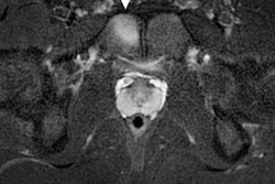
 Professional tennis player with acute onset right groin pain during a match. Axial and coronal fat-saturated T2-weighted MR images demonstrate an acute injury of the right obturator externus muscle with a small focal hematoma. All images courtesy of Dr. Ara Kassarjian.
The lesions he most commonly sees during the tournament are muscles injuries and twisted ankles. The choice of modality is influenced by both context and timing.
"Depending on the timing and type of injury, we may do an ultrasound on the spot or send the patient to the hospital and have him/her come back with the images. I then personally review the imaging results with the whole team," he said. "We work in a shared-decision model with the medical team and the player and his or her coach and manager. It’s really a case-by-case approach."
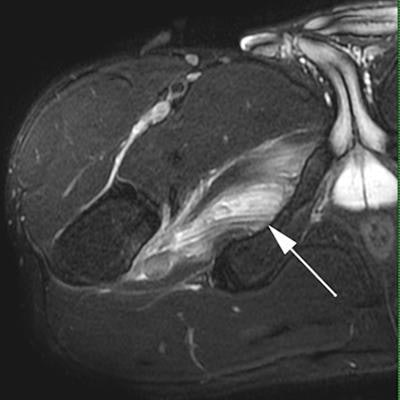
 Professional tennis player who experienced acute pain along the back of his left thigh during a match. Axial and sagittal fat-saturated T2-weighted MR images demonstrate an acute injury to the distal fibers of the long head of the biceps femoris near its junction with the short head biceps femoris.
Tournaments are very intense, with competitions back to back, resulting in a lot of pressure on athletes. "They're peak performers, always on top and under so much internal and external pressure. Pressure is more acute on the younger players now, much more than 10 or 20 years ago," he noted.
Recent advances in both ultrasound and MRI technology have helped diagnose lesions earlier in athletes, allowing for their earlier recovery and return to play.
"We're getting better at MRI and especially at ultrasound of sports injuries," he said. "The sequences, coils, and transducers are getting better and we're learning more as radiologists, we know more about the pathology. Both the technology and medical knowledge of muscle injury have improved over the past few years."
 Professional tennis player who experienced acute onset abdominal wall pain while serving during a match. Transverse ultrasound image of the left rectus abdominus muscle demonstrates an acute muscle injury with edema, acute hemorrhage, and a small focal hyperacute hematoma.
The pandemic and subsequent lockdown in March 2020 shut down sports imaging services in Spain for six weeks. Activity has resumed since then.
Immediately after the first lockdown, Kassarjian noted a slight increase in the number of musculoskeletal injuries in the general population, which has been confirmed by subsequent studies. "We saw a lot of stress fractures and overuse injuries, either because people started to get into shape or, after two months, went immediately back to their previous intensive activity levels. We haven’t noticed as much of an increase in elite athletes," he said.
Kassarjian was actively involved, performing COVID-19 imaging in the emergency setting. Being polyvalent helps him to be better at his job, he believes.
"First, I'm a physician, then a radiologist, then an MSK radiologist. Knowing the rest makes you a better radiologist," he concluded.