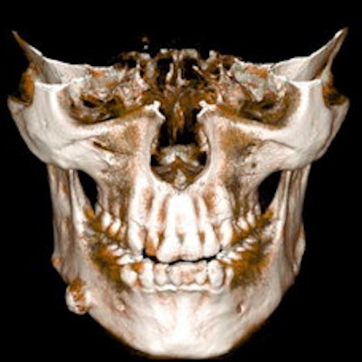
Although clinicians may prefer the photorealistic presentation of cinematic rendering, volume rendering also has discernible benefits in CT scans of the facial skeleton, according to research published online on 9 February in Annals of Anatomy.
A team of researchers from the University of Zurich in Switzerland led by Tobias Steffen had 10 dentists view 10 cases of CT or conebeam CT exams reconstructed with cinematic rendering or volume rendering. After reviewing the images, the dentists subjectively rated the cinematic-rendered images higher.
But volume rendering also may produce images that are sharper and have better contrast.
"Assessing conventional multiplanar CT imaging together with [cinematic rendering] and [volume rendering] reconstructions would give additional, valuable visual information for the surgeon," the authors wrote.
In an exploratory study, the researchers used the same DICOM data to generate both volume-rendering and cinematic-rendering reconstructions. Cinematic rendering was performed using prototype software from Siemens Healthineers, while volume-rendered images were reconstructed using standard clinical postprocessing software (Impax PACS, Agfa HealthCare). To enable an equal comparison, the 10 cases were reconstructed on both techniques using steps of 10-20 degrees.
(A, B) Cinematic rendering reconstruction of an exostosis of the right mandibular corpus, oblique-lateral view. (C, D) Volume rendering reconstruction of the same dataset. Images and caption courtesy of Tobias Steffen et al and Annals of Anatomy through Creative Commons Attribution 4.0 International License.The 10 dentists in the study were asked to view the studies over three different sessions. In the first two sessions, the dentists were randomly shown either a cinematic-rendered image or a volume-rendered image and then asked to fill out a questionnaire rating image characteristics on a visual analog scale from 0 to 10 (best). They then viewed both images together in the last session and again filled out a questionnaire.
The researchers found statistically significant differences in scores for four parameters.
| Dentist scores for images reconstructed with volume vs. cinematic rendering | ||
| Volume rendering | Cinematic rendering | |
| Three-dimensionality | 6.93 | 8.03 |
| Contrast | 5.78 | 8.22 |
| Photorealism | 6.21 | 7.25 |
| Depth perception | 5.87 | 6.83 |
The researchers did not, however, find any significant difference in image interpretation accuracy between the two methods.
Cinematic rendering shows great potential in terms of depth perception and three-dimensionality for CT and CBCT data of the facial skeleton, the researchers concluded. However, volume rendering also offers benefits in surface sharpness and contrast.
"In order to get a first 3D-impression of a pathology, [cinematic rendering] seems highly suitable," the authors wrote. "Therefore, we see [cinematic rendering] as a tool to give additive value to conventional [volume rendering] reconstructions."
The diagnostic value of 3D rotational cinematic-rendered images will need to be assessed in future studies, they said.


![Examples of ultrasound findings and techniques. (A) Images in a 39-year-old male patient with a mass in the left thigh. The mass is heterogeneous on the B-mode US image (compared with the patient in D) and showed increased microvascularity (superb microvascular imaging [SMI]) and shear-wave elastography (SWE) values. Undifferentiated pleomorphic sarcoma was diagnosed at biopsy (with pleomorphic rhabdomyosarcoma in surgical specimen). (B) Images in an 18-year-old male patient with a mass in the left leg. The mass is hypoechoic on the B-mode image, with no other findings suggestive of malignancy. The lesion is in contact with the cortex of the tibia, which is slightly irregular. CT revealed a doubtful anteromedial tibial erosion. The microvascular study demonstrated high vascularization, suggestive of malignancy. Periosteal Ewing sarcoma was diagnosed with both histologic and immunohistochemical confirmation. (C) Images in a 69-year-old female patient with a lump growing on the outside of the left leg. Multiple SWE examinations were performed (please note the high values obtained in the measurements, whereas the color map highlights the stiffness relative to adjacent tissues). SMI showed areas of increased vascularization to target for sampling. Undifferentiated spindle cell sarcoma was diagnosed at biopsy, with residual leiomyosarcoma in the surgical specimen after neoadjuvant therapy. (D) Images in a 56-year-old female patient with a mass in the right thigh. The mass is heterogeneous at both B-mode ultrasound (similar to patient A) and MRI (coronal T2-weighted spectral attenuated inversion recovery [SPAIR]; T1-weighted pre-contrast and postcontrast imaging), which even shows uptake after the administration of paramagnetic contrast material, which is traditionally suggestive of malignancy. Low values at SMI and elastography are suggestive of benignity. Spindle cell lipoma was diagnosed at biopsy, with atypical spindle cell lipomatous tumor in the surgical specimen.](https://img.auntminnieeurope.com/mindful/smg/workspaces/default/uploads/2026/08/images-radiol250278fig2.APCFLSvX6p.jpg?auto=format%2Ccompress&fit=crop&h=100&q=70&w=100)


![Examples of ultrasound findings and techniques. (A) Images in a 39-year-old male patient with a mass in the left thigh. The mass is heterogeneous on the B-mode US image (compared with the patient in D) and showed increased microvascularity (superb microvascular imaging [SMI]) and shear-wave elastography (SWE) values. Undifferentiated pleomorphic sarcoma was diagnosed at biopsy (with pleomorphic rhabdomyosarcoma in surgical specimen). (B) Images in an 18-year-old male patient with a mass in the left leg. The mass is hypoechoic on the B-mode image, with no other findings suggestive of malignancy. The lesion is in contact with the cortex of the tibia, which is slightly irregular. CT revealed a doubtful anteromedial tibial erosion. The microvascular study demonstrated high vascularization, suggestive of malignancy. Periosteal Ewing sarcoma was diagnosed with both histologic and immunohistochemical confirmation. (C) Images in a 69-year-old female patient with a lump growing on the outside of the left leg. Multiple SWE examinations were performed (please note the high values obtained in the measurements, whereas the color map highlights the stiffness relative to adjacent tissues). SMI showed areas of increased vascularization to target for sampling. Undifferentiated spindle cell sarcoma was diagnosed at biopsy, with residual leiomyosarcoma in the surgical specimen after neoadjuvant therapy. (D) Images in a 56-year-old female patient with a mass in the right thigh. The mass is heterogeneous at both B-mode ultrasound (similar to patient A) and MRI (coronal T2-weighted spectral attenuated inversion recovery [SPAIR]; T1-weighted pre-contrast and postcontrast imaging), which even shows uptake after the administration of paramagnetic contrast material, which is traditionally suggestive of malignancy. Low values at SMI and elastography are suggestive of benignity. Spindle cell lipoma was diagnosed at biopsy, with atypical spindle cell lipomatous tumor in the surgical specimen.](https://img.auntminnieeurope.com/mindful/smg/workspaces/default/uploads/2026/08/images-radiol250278fig2.APCFLSvX6p.jpg?auto=format%2Ccompress&fit=crop&h=112&q=70&w=112)