The radiology training program at a major tertiary hospital in Sydney has lost its accreditation, according to a report posted on 11 August by the Sydney Morning Herald (SMH). The move raises fears that other programs might face a similar fate.
The Royal Australian and New Zealand College of Radiologists (RANZCR) decided the program of the 975-bed Westmead Hospital failed to meet its standards. The scheme supports 15 trainee radiologists, and the accreditation will expire on 17 September, the SMH noted.
"There is inadequate supervision of training in the radiology department at Westmead Hospital and trainees are not getting the necessary teaching and education," said Dr. Lance Lawler, president of the RANZCR, adding that his team is happy to work with Westmead Hospital to help reestablish accreditation.
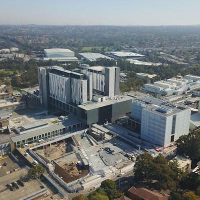
This aerial photo of the Westmead shows the new central acute services building, which includes two new emergency departments and 300 patient rooms and is part of a 1-billion Australian dollar transformation of the Westmead Health Precinct in Western Sydney. Photo courtesy of Xinhua/Alamy Stock PhotoThe decision comes following a 2019 review by the RANZCR, after which the Westmead received a D grade, the lowest level. The department was placed "on watch" with an opportunity to improve, but that did not happen.
According to the SMH, sources who are familiar with the situation but not authorized to comment publicly said the latest RANZCR decision was not influenced by staff furloughing due to recent COVID-19 exposures.
Staff have reportedly expressed concern about whether the hospital can continue to offer 24-hour radiology services without trainees. An email sent by the hospital's general manager to staff, seen by the SMH, suggested the loss of the trainee workforce could have a "profound impact" on the availability of radiology services at the hospital, particularly after hours.
"The radiology department at Westmead remains fully operational and appropriately staffed, and our patients will continue to receive the highest quality radiology services," the district health service said in a statement. "We are supporting those radiology trainees affected by the College's decision to withdraw its teaching accreditation."
Serious situation
Australian Medical Association president Dr. Danielle McMullen described the situation as "very serious," the SMH reported.
"We are in discussion with the parties involved and encourage Westmead and the local health district to meet the required standards as soon as possible to reestablish accreditation and provide a safe, supportive training environment for registrars," she said. "Providing high-quality patient care will continue to be a priority for the clinicians working at the hospital."
One of the issues that can occur with accreditation is excessive workload/volume versus RANZCR workload standards for accreditation, a source told AuntMinnieEurope.com.
As is the case in every other country, radiology workload continues to increase more than staffing levels in Australia. Unlike the U.K. Royal College of Radiologists, for instance, the RANZCR has workload standards for training departments but not for safe workload in general, the source said.

![Examples of ultrasound findings and techniques. (A) Images in a 39-year-old male patient with a mass in the left thigh. The mass is heterogeneous on the B-mode US image (compared with the patient in D) and showed increased microvascularity (superb microvascular imaging [SMI]) and shear-wave elastography (SWE) values. Undifferentiated pleomorphic sarcoma was diagnosed at biopsy (with pleomorphic rhabdomyosarcoma in surgical specimen). (B) Images in an 18-year-old male patient with a mass in the left leg. The mass is hypoechoic on the B-mode image, with no other findings suggestive of malignancy. The lesion is in contact with the cortex of the tibia, which is slightly irregular. CT revealed a doubtful anteromedial tibial erosion. The microvascular study demonstrated high vascularization, suggestive of malignancy. Periosteal Ewing sarcoma was diagnosed with both histologic and immunohistochemical confirmation. (C) Images in a 69-year-old female patient with a lump growing on the outside of the left leg. Multiple SWE examinations were performed (please note the high values obtained in the measurements, whereas the color map highlights the stiffness relative to adjacent tissues). SMI showed areas of increased vascularization to target for sampling. Undifferentiated spindle cell sarcoma was diagnosed at biopsy, with residual leiomyosarcoma in the surgical specimen after neoadjuvant therapy. (D) Images in a 56-year-old female patient with a mass in the right thigh. The mass is heterogeneous at both B-mode ultrasound (similar to patient A) and MRI (coronal T2-weighted spectral attenuated inversion recovery [SPAIR]; T1-weighted pre-contrast and postcontrast imaging), which even shows uptake after the administration of paramagnetic contrast material, which is traditionally suggestive of malignancy. Low values at SMI and elastography are suggestive of benignity. Spindle cell lipoma was diagnosed at biopsy, with atypical spindle cell lipomatous tumor in the surgical specimen.](https://img.auntminnieeurope.com/mindful/smg/workspaces/default/uploads/2026/08/images-radiol250278fig2.APCFLSvX6p.jpg?auto=format%2Ccompress&fit=crop&h=100&q=70&w=100)


![Examples of ultrasound findings and techniques. (A) Images in a 39-year-old male patient with a mass in the left thigh. The mass is heterogeneous on the B-mode US image (compared with the patient in D) and showed increased microvascularity (superb microvascular imaging [SMI]) and shear-wave elastography (SWE) values. Undifferentiated pleomorphic sarcoma was diagnosed at biopsy (with pleomorphic rhabdomyosarcoma in surgical specimen). (B) Images in an 18-year-old male patient with a mass in the left leg. The mass is hypoechoic on the B-mode image, with no other findings suggestive of malignancy. The lesion is in contact with the cortex of the tibia, which is slightly irregular. CT revealed a doubtful anteromedial tibial erosion. The microvascular study demonstrated high vascularization, suggestive of malignancy. Periosteal Ewing sarcoma was diagnosed with both histologic and immunohistochemical confirmation. (C) Images in a 69-year-old female patient with a lump growing on the outside of the left leg. Multiple SWE examinations were performed (please note the high values obtained in the measurements, whereas the color map highlights the stiffness relative to adjacent tissues). SMI showed areas of increased vascularization to target for sampling. Undifferentiated spindle cell sarcoma was diagnosed at biopsy, with residual leiomyosarcoma in the surgical specimen after neoadjuvant therapy. (D) Images in a 56-year-old female patient with a mass in the right thigh. The mass is heterogeneous at both B-mode ultrasound (similar to patient A) and MRI (coronal T2-weighted spectral attenuated inversion recovery [SPAIR]; T1-weighted pre-contrast and postcontrast imaging), which even shows uptake after the administration of paramagnetic contrast material, which is traditionally suggestive of malignancy. Low values at SMI and elastography are suggestive of benignity. Spindle cell lipoma was diagnosed at biopsy, with atypical spindle cell lipomatous tumor in the surgical specimen.](https://img.auntminnieeurope.com/mindful/smg/workspaces/default/uploads/2026/08/images-radiol250278fig2.APCFLSvX6p.jpg?auto=format%2Ccompress&fit=crop&h=112&q=70&w=112)