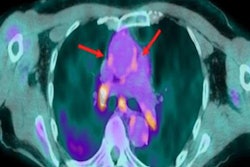
The number of scintigraphy studies -- particularly thyroid, cardiac, bone, and lung scans -- fell sharply during the first year of the COVID-19 pandemic, while PET scans have remained stable, a large-scale European survey has found.
"Benign therapy was more severely affected overall than malignant therapy," wrote first author Dr. Ana Paula Moreira, a nuclear medicine physician at Coimbra University Hospital and the Institute for Nuclear Sciences Applied to Health (ICNAS) at Coimbra University in Portugal, and colleagues. "Despite all the difficulties, there has been an effort to prioritize the assessment of cancer patients."
 Dr. Ana Paula Moreira.
All 39 European Association of Nuclear Medicine (EANM) national delegates were asked to complete the survey in January 2021, and 32 of them did so. The survey focused on the pandemic's impact on diagnostic (scintigraphy and PET) and therapeutic nuclear medicine procedures, supply of radionuclides or radiopharmaceuticals, and operational aspects of nuclear medicine departments.
The full results were posted on 30 July in the European Journal of Nuclear Medicine and Molecular Imaging.
Overall, the number of scans fell by between 25% and 50% for most benign therapies. Diagnostic PET/CT scans and malignant therapeutic procedures remained relatively stable in most of Europe, with rates of 69% and 66%, respectively. The problem of secure radiopharmaceutical supply was only a factor for 25% of the respondents.
PET/CT was less severely affected because local cyclotron centers can produce PET radionuclides and tracers, whereas some countries rely on regular international flights for their weekly supply of generators (e.g., molybdenum-99/technetium-99m) and some radionuclides (iodine-131), the authors speculated.
"There was even an increase in PET/CT procedures in two countries," they stated. "One explanation for this might be the overbooked CT capacities, which resulted in PET/CT becoming a one-stop staging tool without prior CT."
The most severely affected months were March, April, and May 2020, probably due to the sudden outbreak of the disease and the unexpected need for departmental reorganization, according to the authors. Some countries reported a lower number of exams during spring 2020, but a subsequent increase in autumn 2020, resulting in a stable situation for the whole year.
Workflow and training
Organization/management workflows underwent changes in two-thirds of European nuclear medicine departments, the survey found. Workflow depended on the official and legally imposed lockdown periods, mainly implemented not only to reduce the circulation of the virus but also to provide staff for the most urgent tasks, such as redeployment of nursing staff and physicians to COVID-19 units.
"The reduced contact with physicians in outpatient consultations in the initial period of the pandemic resulted in a decreased number of patients being referred to nuclear medicine for diagnosis and treatment, this effect being less prominent for oncological patients," wrote Moreira and colleagues.
Another factor was patient no-shows due to the fear of being infected at the hospital or using public transport to reach hospitals, but this was not quantified at the level of the EANM member countries.
"The pandemic probably not only affected healthcare services but also the training and education of our residents and young fellows, the future generation of nuclear medicine physicians," they noted. "Our residents were able to attend a smaller number of studies/courses than in previous years. Some of them were even assigned to rotations for COVID-19 services for several months, hence academic productivity and research projects in the field of nuclear medicine might also have been affected."
The survey responses were completed before the "third wave" was apparent, and this may have changed the situation, the authors wrote.
Unanswered questions
To provide follow-up perspective, the EANM is planning a repeat survey in 2022.
"There are still some unanswered questions," Moreira told AuntMinnieEurope.com. "Undoubtedly one of them is the role of nuclear medicine in the study of COVID-19 complications and sequelae. I imagine that well-known radiopharmaceuticals and procedures like F-18 FDG PET/CT, radiolabeled white blood cells (scintigraphy, SPECT, SPECT/CT), or even lung ventilation/perfusion SPECT may be useful in this new clinical scenario."
Because nuclear medicine techniques have inherent potential in new drug development, it can be expected in the not so distant future that the development of new radiopharmaceuticals will contribute to diagnostic, prognostic, and even therapeutic tools for SARS-CoV-2 infection, she said.
"Next year, I’m sure that nuclear medicine departments will have less difficulties facing the pandemic, since the adaptation period to this public health crisis has already been overcome, in most cases with great success," Moreira concluded.