Dear AuntMinnieEurope Member,
The rising incidence of renal pathologies also means a growing incidence of complications from renal interventional procedures, according to the story that was the most-viewed article on AuntMinnieEurope.com this past week.
A group from Australia discussed the role of radiologists in detecting possible complications from renal interventions, especially with the growing shift to less invasive procedures. Multiple imaging modalities can help, including CT, MRI, and digital subtraction angiography.
In addition to causing tragic loss of life over the past year, the COVID-19 pandemic has upended society in myriad ways. For example, many radiologist board certification exams had to be postponed and rescheduled -- including the European Diploma in Radiology (EDiR). In a new article, Prof. Laura Oleaga of the European Board of Radiology (EBR) discussed how the EBR has adapted.
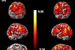
The pandemic has also caused anxiety and workplace-related stress among radiographers, according to a new study out of the U.K. Established working patterns and workloads have changed, with many radiographers not receiving the training they need to care for patients with COVID-19.
In other news, Swiss researchers found a unique sign on radiography indicating a painful foot neuroma -- a V-shaped sign that they named after the character Mr. Spock from "Star Trek." And cardiac imaging research in Spain is set to get a big boost thanks to a new scholarship program.


![Overview of the study design. (A) The fully automated deep learning framework was developed to estimate body composition (BC) (defined as subcutaneous adipose tissue [SAT] in liters; visceral adipose tissue [VAT] in liters; skeletal muscle [SM] in liters; SM fat fraction [SMFF] as a percentage; and intramuscular adipose tissue [IMAT] in deciliters) from MRI. The fully automated framework comprised one model (model 1) to quantify different BC measures (SAT, VAT, SM, SMFF, and IMAT) as three-dimensional (3D) measures from whole-body MRI scans. The second model (model 2) was trained to identify standardized anatomic landmarks along the craniocaudal body axis (z coordinate field), which allowed for subdividing the whole-body measures into different subregions typically examined on clinical routine MRI scans (chest, abdomen, and pelvis). (B) BC was quantified from whole-body MRI in over 66,000 individuals from two large population-based cohort studies, the UK Biobank (UKB) (36,317 individuals) and the German National Cohort (NAKO) (30,291 individuals). Bar graphs show age distribution by sex and cohort. BMI = body mass index. (C) After the performance assessment of the fully automated framework, the change in BC measures, distributions, and profiles across age decades were investigated. Age-, sex-, and height-adjusted body composition reference curves were calculated and made publicly available in a web-based z-score calculator (https://circ-ml.github.io).](https://img.auntminnieeurope.com/mindful/smg/workspaces/default/uploads/2026/05/body-comp.XgAjTfPj1W.jpg?auto=format%2Ccompress&fit=crop&h=112&q=70&w=112)


