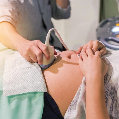
A lively debate is underway in the U.K. over whether a woman's partner should be allowed to sit in on her obstetric ultrasound examination. Speculation is growing that patients in England might soon have a legal right to insist on bringing a support person to all appointments in the maternity pathway.
The Society of Radiographers (SoR) is urging caution. In a BBC Radio 4 Women's Hour interview on 26 March (from 30.12 to 37 mins into the recording), Gill Harrison, the society's professional officer for ultrasound, acknowledged that support people are important when a woman undergoes obstetric ultrasound, but she highlighted the need to keep other patients, as well as sonographers, safe from COVID-19.
Having an extra person in the department may put other patients and the sonographer at risk for contracting the disease, increases exam prep and clean-up time, and it may distract the sonographer from the task, she said. Harrison has deep concerns that live streaming or recording of the exam by a partner would have a serious impact on sonographer concentration whilst performing a complex clinical examination.
According to a statement posted on the SoR website on 29 March, sonographers across England are reporting rumors of a directive expected from the Department of Health and Social Care regarding the expectation that a support person must be able to attend for all appointments in the maternity pathway. This would include all antenatal ultrasound appointments.
"No official policy change has been released. SoR is engaged with members of the Chief Allied Health Professions Officer team at NHS England and will keep members informed as soon as we hear about any change," the statement noted.
The BBC interview was prompted in part by an opinion article written by London sonographer Anna Mada and published in Synergy News in February called "We cannot allow obstetric ultrasound to become entertainment."