CT has offered fresh clues about the killing of a significant pharaoh who ruled Egypt around 3,600 years ago. Experts now believe Seqenenre Taa II -- known as "the Brave" -- was killed in an execution ceremony after being taken prisoner on the battlefield.
Seqenenre reigned over southern Egypt, and he led troops against the Hyksos, a dynasty of West Asian origin that had taken over the Nile Delta. His mummy was examined in the 1960s, and x-ray revealed head wounds concealed by embalmers, giving rise to theories that he was killed in battle or a palace assassination.
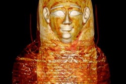
 Coronal CT image of the head and neck of the mummy of Seqenenre Taa II. The desiccated brain is seen shifted and occupying the left side of the cranial cavity. All images courtesy of Dr. Sahar Saleem and Frontiers in Medicine.
Dr. Sahar Saleem, a professor of radiology at Cairo University who specializes in CT mummy imaging, and archeologist Zahi Hawass, PhD, have used CT scans and 3D reconstructions to suggest he had multiple killers. They have presented their findings in an article published on 17 February in Frontiers of Medicine that received over 10,500 page views by 21 February.
CT revealed details of the head injuries, including wounds that had not been discovered in previous examinations and had been skillfully hidden by the embalmers. The mummy's deformed hands indicate that Seqenenre may have been captured on the battlefield, and his hands were tied behind his back, preventing him from deflecting the fierce attack on his head.
 Coronal 3D CT image of lower torso of Seqenenre shows evisceration and embalming material within the abdomino-pelvic cavity. Note the disarticulated vertebrae and disrupted symphyseal articulation.
"This suggests that Seqenenre was really on the front line with his soldiers risking his life to liberate Egypt," said Saleem. "In a normal execution on a bound prisoner, it could be assumed that only one assailant strikes, possibly from different angles but not with different weapons."
The CT scans, combined with other evidence, suggest the execution was carried out by multiple attackers, which the scientists confirmed by studying five different Hyksos weapons that matched the king's wounds. The study also determined that Seqenenre was about 40 when he died, based on the detailed morphology revealed in the images, providing the most precise estimate to date, according to the authors.
 3D frontal CT image of upper limbs of Seqenenre. The arms are dislocated at the shoulder and bent at the elbows. Note the deformity of the bent hands, indicating they were tied at the wrists.
In addition, the investigation revealed details about the mummification of Seqenenre's body. For instance, the embalmers used a sophisticated method to hide the king's head wounds under a layer of embalming material that functioned similarly to the fillers used in modern plastic surgery. This would imply that mummification took place in a real mummification laboratory rather than in a poorly equipped place, as previously interpreted.
Saleem said the study provides important new details about a pivotal point in Egypt's long history. Seqenenre's death motivated his successors to continue the fight to unify Egypt and start the New Kingdom, she said.
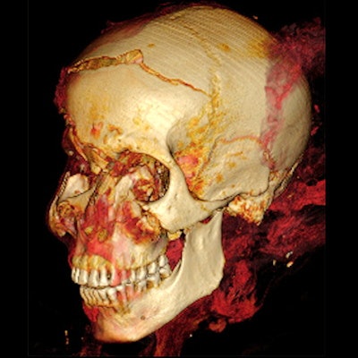
 3D CT image of head of Seqenenre in frontal projection showing multiple craniofacial injuries: a gaping fracture of the frontal bone; fracture above the right eyebrow; comminuted fractures of the nose, right orbit, and right zygoma caused by blunt trauma; and a small perforating wound overlying the right cheek caused by fragments of the fractured zygoma.
Researchers have spent decades trying to decipher the death of Seqenenre, whose body was found in the late 19th century and had visible wounds on its face.
Royal mummies in Cairo
Saleem examined the mummy on 4 May 2019. She said she used the usual optimum CT scan parameters, including field-of-view and slice thickness, to provide the best 3D reconstruction. Because of the COVID-19 pandemic, it took several weeks to study the Hyksos weapons stored at the Cairo Egyptian Museum and to correlate them with the injuries by physical inspection of the mummy and on CT scans.
 3D CT image of Seqenenre's head in left oblique projection shows an oblique cut wound of the left zygoma and fracture of left coronoid process of the mandible.
She has conducted CT scans for all the royal mummies housed at the Egyptian Museum in Cairo, using the CT machine located in the garden of the museum. She recently investigated 15 royal mummies that have not been scanned before with CT, including Seti II, Amenhotep II, Siptah, and Ramesses IV, V, VI, and IX.
Soon, the royal mummies will be transferred from the Egyptian Museum to the National Museum of Egyptian Civilization (NMEC), a new facility in Cairo. To avoid unnecessary transportation of the mummies, it was convenient to do the scans using the CT unit at the Egyptian Museum while they were still housed there.
 Axial thick slab 3D reconstructed CT image of the external view of the skull base of Seqenenre's shows fracture of the left lip of foramen magnum inflicted by a penetrating injury of the left mastoid bone.
"The results of the investigations helped in preparing the mummies to be transferred to the new museum. The Egyptian Ministry of Antiquities appointed me as a member of the NMEC's Display Scenario committee," Saleem told AuntMinnieEurope.com. "I used illustrated CT scan images to be displayed alongside some of the royal mummies in their new home. Digital illustrations will enhance the museum visitors' experience."