A PET-based optical imaging technique called Cerenkov luminescence imaging (CLI) was successfully used by German researchers to confirm the presence of cancer cells after prostate cancer surgery, according to a study published in the October edition of the Journal of Nuclear Medicine.
Researchers from the University Medical Center Essen tested the use of Cerenkov luminescence imaging in a small group of 10 patients with high-risk primary prostate cancer (Journal of Nuclear Medicine, October 2020, Vol. 61:10, pp. 1500-1506). The goal of the study authors was to see if CLI could accurately detect surgical margins during radical prostatectomy and to indicate whether residual cancer remained after the surgery.
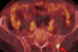
Cerenkov luminescence imaging is a phenomenon in which optical photons are emitted by PET radiotracers that can be detected in addition to the positrons that are used for conventional PET imaging. In the case of this study, patients were imaged with a gallium-68 (Ga-68) prostate-specific membrane antigen (PSMA) radiotracer.
Under the Essen group's protocol, the Ga-68 PSMA PET scans were performed first, followed by the radical prostatectomy procedure. Next, intraoperative CLI was performed on the excised prostate. Readers then analyzed the CLI images; signal intensity and tumor-to-background ratios were calculated for different regions of interest.
The researchers calculated tumor margins with CLI by analyzing elevated signals at the surface of intact prostate images. To validate the CLI technique's accuracy, the margin status assessments were compared with histology performed after surgery.
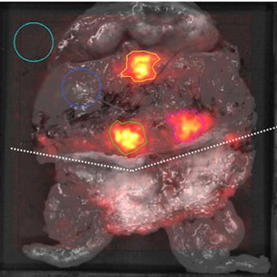
 Grayscale photographic images overlaid with Cerenkov luminescence imaging signals. Incised prostate gland specimen of patient number 1 (A) and 2 (B). In each image, the regions of interest (ROIs) are encircled. The incision is marked with a dotted line. The light blue ROI (area of empty specimen tray) and dark blue ROI (area of normal prostate tissue) were used as empty background and tissue background, respectively. The green ROI (lesion 1) and pink ROI (lesion 2) show an increased signal; histopathological analysis confirmed cancer tissue in these areas. The orange ROI in image B tshows an increased signal from an area without cancer cells. Images created by Christopher Darr, PhD, and Dr. Ina Binse, University Medical Center Essen. Images courtesy of the Society of Nuclear Medicine and Molecular Imaging (SNMMI).
The researchers found that readers were able to detect tumor cells on the Cerenkov luminescence images -- a finding that was confirmed with histology. Three of the 10 patients had positive surgical margins, and two had elevated signal levels on the CLI images. In all, CLI correctly indicated tumor signal in 25 out of 35 regions of interest on the optical images.
The researchers concluded that intraoperative Cerenkov luminescence imaging could improve the accuracy and safety of radical prostatectomy, especially in high-risk prostate cancer patients. They also suggested that targeted surgical removal of lymph node metastases could be performed with CLI.