
The Spanish Society of Medical Radiology (SERAM) has posted a tribute to Dr. Cristina Corbella Sala, a neuroradiologist at Mútua Terrassa University Hospital, near Barcelona. She died recently in her early 50s.
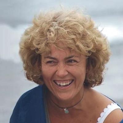
Dr. Cristina Corbella Sala.Corbella Sala died of amyotrophic lateral sclerosis, also known as Lou Gehrig's disease. She faced the disease with dignity, participating in forums that raised awareness of the disease, sharing her experiences of the illness, and raising funds for charities, according to the SERAM tribute.
"She was an optimistic and cheerful person and that is how she has been until the end," Prof. Laura Oleaga, PhD, chair of radiology at the Hospital Clinic of Barcelona, told AuntMinnieEurope.com. "Within radiology she always had concerns to progress and transmit her knowledge to young people. She was a great radiologist and an excellent person."
She loved nature and particularly enjoyed the mountains. "I have seen a video of her sliding down the snow slopes in an adapted chair driven by a family member this past winter," Oleaga recalls.
Corbella Sala will be remembered for her passion for medicine, radiology, and neuroradiology and for being an expert in the field of head and neck pathology, and will also be remembered for her devotion to her children, noted SERAM.


![Overview of the study design. (A) The fully automated deep learning framework was developed to estimate body composition (BC) (defined as subcutaneous adipose tissue [SAT] in liters; visceral adipose tissue [VAT] in liters; skeletal muscle [SM] in liters; SM fat fraction [SMFF] as a percentage; and intramuscular adipose tissue [IMAT] in deciliters) from MRI. The fully automated framework comprised one model (model 1) to quantify different BC measures (SAT, VAT, SM, SMFF, and IMAT) as three-dimensional (3D) measures from whole-body MRI scans. The second model (model 2) was trained to identify standardized anatomic landmarks along the craniocaudal body axis (z coordinate field), which allowed for subdividing the whole-body measures into different subregions typically examined on clinical routine MRI scans (chest, abdomen, and pelvis). (B) BC was quantified from whole-body MRI in over 66,000 individuals from two large population-based cohort studies, the UK Biobank (UKB) (36,317 individuals) and the German National Cohort (NAKO) (30,291 individuals). Bar graphs show age distribution by sex and cohort. BMI = body mass index. (C) After the performance assessment of the fully automated framework, the change in BC measures, distributions, and profiles across age decades were investigated. Age-, sex-, and height-adjusted body composition reference curves were calculated and made publicly available in a web-based z-score calculator (https://circ-ml.github.io).](https://img.auntminnieeurope.com/mindful/smg/workspaces/default/uploads/2026/05/body-comp.XgAjTfPj1W.jpg?auto=format%2Ccompress&fit=crop&h=112&q=70&w=112)


