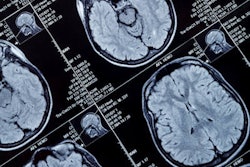
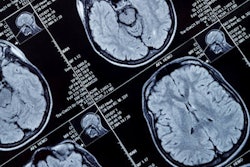
U.K. researchers are offering an outline of neurological manifestations of severe SARS-CoV-2 infection they hope will help their colleagues better recognize and treat COVID-19, the disease caused by the virus, according to a study published on 8 July in Brain.
The neurological complications of COVID-19 are becoming more obvious as the pandemic wears on, and they consist of a startling range of conditions, according to a team led by Ross Paterson, PhD, of University College London.
"The widespread effects of COVID-19 include neurological disorders, but there have been, to date, no detailed clinical reports of their nature," the group wrote. "Our London and regional cohort describe a range of neurological symptoms including encephalopathies, para- and postinfectious central nervous system syndromes, including encephalitis, [acute disseminated encephalomyelitis] with hemorrhage and necrotic change, transverse myelitis, ischemic stroke and Guillain-Barré syndrome."
To develop an understanding of the emerging spectrum of neurological manifestations of COVID-19, Paterson and colleagues used data from 43 patients who were referred to London's National Hospital, Queen Square COVID-19 multidisciplinary team. The authors reviewed clinical, radiological, laboratory, and neuropathological findings from these patients between April 9 and May 15.
Of the 43 patients, 29 were positive for SARS-CoV-2, eight were probably positive, and six were possibly infected. Half of the patients (53%) were people of color. The brain imaging patients underwent included both MRI and CT exams.
The team discovered five categories of neurological manifestations:
| Neurological manifestations of COVID-19 | ||||
| Type of neurological manifestation (number of patients) | Median patient age | Days of COVID-19 infection before neurological symptoms | Treatment | Outcome |
| Encephalopathy (10), with delirium/psychosis and no distinct MRI or CSF abnormalities | 57.5 | 4.5 | 9 treated with supportive measures, 1 with steroids | 7 complete recoveries, 2 partial |
| Inflammatory central nervous system syndromes (12) | 53 | 9 | 10 treated with corticosteroids; 3 with intravenous immunoglobulin | 1 complete recovery, 10 partial, 1 death |
| Stroke (8) | 62.5 | 8 | 7 with low molecular weight heparin; 1 with apixaban | 7 incomplete recoveries, 1 death |
| Peripheral syndromes (8) | ||||
| Guillain-Barré syndrome (7) | 57 | 13 | 7 with intravenous immunoglobulin | 7 incomplete recoveries |
| Plexopathy (1) | 60 | 14 | Intravenous steroids | Incomplete recovery |
| Miscellaneous/uncharacterized (5) | 20 | 10 | All 5 treated with steroids | 1 complete recovery, 3 partial, 1 "nil" |
SARS-CoV-2 infection appears to manifest in a variety of neurological syndromes that can affect the whole nervous system, the group wrote.
"The high incidence of acute disseminated encephalomyelitis, particularly with hemorrhagic change, is striking," the authors wrote. "This complication was not related to the severity of the respiratory COVID-19 disease."
More research is definitely needed regarding neurological complications of SARS-CoV-2 infection, according to Paterson and colleagues.
"Further clinical, neuroradiological, biomarker, and neuropathological studies are essential to determine the underlying pathobiological mechanisms, which will guide treatment," they concluded. "Longitudinal follow-up studies will be necessary to ascertain the long-term neurological and neuropsychological consequences of this pandemic."


![Examples of ultrasound findings and techniques. (A) Images in a 39-year-old male patient with a mass in the left thigh. The mass is heterogeneous on the B-mode US image (compared with the patient in D) and showed increased microvascularity (superb microvascular imaging [SMI]) and shear-wave elastography (SWE) values. Undifferentiated pleomorphic sarcoma was diagnosed at biopsy (with pleomorphic rhabdomyosarcoma in surgical specimen). (B) Images in an 18-year-old male patient with a mass in the left leg. The mass is hypoechoic on the B-mode image, with no other findings suggestive of malignancy. The lesion is in contact with the cortex of the tibia, which is slightly irregular. CT revealed a doubtful anteromedial tibial erosion. The microvascular study demonstrated high vascularization, suggestive of malignancy. Periosteal Ewing sarcoma was diagnosed with both histologic and immunohistochemical confirmation. (C) Images in a 69-year-old female patient with a lump growing on the outside of the left leg. Multiple SWE examinations were performed (please note the high values obtained in the measurements, whereas the color map highlights the stiffness relative to adjacent tissues). SMI showed areas of increased vascularization to target for sampling. Undifferentiated spindle cell sarcoma was diagnosed at biopsy, with residual leiomyosarcoma in the surgical specimen after neoadjuvant therapy. (D) Images in a 56-year-old female patient with a mass in the right thigh. The mass is heterogeneous at both B-mode ultrasound (similar to patient A) and MRI (coronal T2-weighted spectral attenuated inversion recovery [SPAIR]; T1-weighted pre-contrast and postcontrast imaging), which even shows uptake after the administration of paramagnetic contrast material, which is traditionally suggestive of malignancy. Low values at SMI and elastography are suggestive of benignity. Spindle cell lipoma was diagnosed at biopsy, with atypical spindle cell lipomatous tumor in the surgical specimen.](https://img.auntminnieeurope.com/mindful/smg/workspaces/default/uploads/2026/08/images-radiol250278fig2.APCFLSvX6p.jpg?auto=format%2Ccompress&fit=crop&h=100&q=70&w=100)


![Examples of ultrasound findings and techniques. (A) Images in a 39-year-old male patient with a mass in the left thigh. The mass is heterogeneous on the B-mode US image (compared with the patient in D) and showed increased microvascularity (superb microvascular imaging [SMI]) and shear-wave elastography (SWE) values. Undifferentiated pleomorphic sarcoma was diagnosed at biopsy (with pleomorphic rhabdomyosarcoma in surgical specimen). (B) Images in an 18-year-old male patient with a mass in the left leg. The mass is hypoechoic on the B-mode image, with no other findings suggestive of malignancy. The lesion is in contact with the cortex of the tibia, which is slightly irregular. CT revealed a doubtful anteromedial tibial erosion. The microvascular study demonstrated high vascularization, suggestive of malignancy. Periosteal Ewing sarcoma was diagnosed with both histologic and immunohistochemical confirmation. (C) Images in a 69-year-old female patient with a lump growing on the outside of the left leg. Multiple SWE examinations were performed (please note the high values obtained in the measurements, whereas the color map highlights the stiffness relative to adjacent tissues). SMI showed areas of increased vascularization to target for sampling. Undifferentiated spindle cell sarcoma was diagnosed at biopsy, with residual leiomyosarcoma in the surgical specimen after neoadjuvant therapy. (D) Images in a 56-year-old female patient with a mass in the right thigh. The mass is heterogeneous at both B-mode ultrasound (similar to patient A) and MRI (coronal T2-weighted spectral attenuated inversion recovery [SPAIR]; T1-weighted pre-contrast and postcontrast imaging), which even shows uptake after the administration of paramagnetic contrast material, which is traditionally suggestive of malignancy. Low values at SMI and elastography are suggestive of benignity. Spindle cell lipoma was diagnosed at biopsy, with atypical spindle cell lipomatous tumor in the surgical specimen.](https://img.auntminnieeurope.com/mindful/smg/workspaces/default/uploads/2026/08/images-radiol250278fig2.APCFLSvX6p.jpg?auto=format%2Ccompress&fit=crop&h=112&q=70&w=112)