If there is one positive thing to come out of the COVID-19 pandemic, it is the insight that humanity can only solve today's complex problems through international collaboration. That's the central message of a group of radiologists who wrote an article published on 8 April in European Radiology, in which Dr. Liang Wang, vice chairman for clinical affair in the radiology department at Tongji Hospital in Wuhan, China, shares his facility's strategies and experiences.
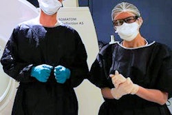
Among the recommendations from Wuhan is taking steps to avoid cross-infections. The precautions include dividing workplaces into "clear different zones" to ensure distance between clinicians and radiologists and exiting areas with potentially infected patients only after completing cleaning and disinfection procedures.
In addition, the Wuhan facility required both x-ray technicians and patients to wear protective equipment, such as face masks, during the examination. The staff also treated used supplies from COVID-19-related imaging procedures as medical waste, packing the leftovers in "double yellow medical waste bags" and marking them as "infectious COVID-19 waste" before final disposal.
To what degree strategies used by Chinese radiologists and healthcare practitioners can be used effectively in Europe is debatable, but the guidance is helpful in preventing the spread of the novel coronavirus, the authors wrote.
"We expect an enormous increase of COVID-19 patients in the next weeks and months all over the world and also in Europe," they cautioned. "If radiology departments want to think about whether their own plans are sufficient, they can compare them with the procedures in Wuhan. We hope this will give our European colleagues the opportunity to learn how a national crisis has been successfully managed."