MRI scans have revealed changes in the brain structure of individuals who continually exhibit antisocial behavior, according to a study conducted at a top London facility and published in the March issue of Lancet Psychiatry.
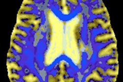
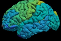
The MR images showed a thinner cortex and smaller surface area in regions of the brain that could be linked to a person's tendency to steal, bully, and act aggressively and irresponsibly.
"Our findings support the idea that, for the small proportion of individuals with life-course-persistent antisocial behavior, there may be differences in their brain structure that make it difficult for them to develop social skills that prevent them from engaging in antisocial behavior," said lead author Christina Carlisi, PhD, from University College London (UCL) in a statement. "These people could benefit from more support throughout their lives."
Carlisi and colleagues analyzed brain MRI scans from 672 participants (mean age, 45 years), along with behavioral reports from parents, teachers, and the subjects themselves on conduct issues between ages 7 and 26 years. The individuals were divided into three groups: 80 people (12%) with life-course-persistent antisocial behavior, 151 (23%) with adolescent-only antisocial behavior, and 441 (66%) with no history of persistent antisocial behavior (Lancet Psychiat, March 2020, Vol. 7:3, pp. 245-253).
Compared with people with no persistent antisocial behavior, those with life-course-persistent antisocial behavior had reduced surface area in 282 (78%) of 360 brain regions and a thinner cortex in 11 (3%) of 360 regions.
However, the researchers found no widespread differences in brain structure between the group with behavior issues during adolescence only and the normal group or the life-course-persistent antisocial group.
"Most people who exhibit antisocial behavior primarily do so only in adolescence, likely as a result of navigating socially difficult years, and these individuals do not display structural brain differences," Carlisi added. "It is also these individuals who are generally capable of reform and go on to become valuable members of society."
The authors recommended more long-term studies of antisocial behavior, measuring brain changes, genes, and a person's environment, to better understand how life-course-persistent antisocial behavior unfolds.
"It is unclear whether these brain differences are inherited and precede antisocial behavior, or whether they are the result of a lifetime of confounding risk factors (e.g., substance abuse, low IQ, and mental health problems) and are therefore a consequence of a persistently antisocial lifestyle," pointed out co-author Prof. Essi Viding, also from UCL.
Carlisi works as a Wellcome Trust-funded Sir Henry Wellcome postdoctoral fellow at UCL. Her research in the Developmental Risk and Resilience Unit uses structural equation modeling and computational methods in multidimensional data to examine individual differences in the mechanisms of affective processing, and she is involved in developing more sensitive measures of these differences to study how psychiatric traits develop across adolescence.
In her doctoral program, completed in 2017 at King's College London, she used functional MRI to study shared and disorder-specific neurofunctional abnormalities related to executive function in adolescents with autism spectrum disorder and obsessive-compulsive disorder.