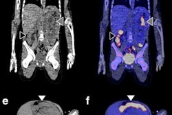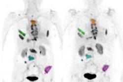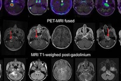
Researchers are one step closer to proving the worth of zirconium-89 (Zr-89) immuno-PET as a noninvasive approach to predicting the effectiveness of therapy for cancerous tumors, according to a study published in the December issue of the Journal of Nuclear Medicine.
The proof-of-concept results show that Zr-89 immuno-PET can be used to measure monoclonal antibody concentrations in normal tissues and, by doing so, potentially lead to more personalized, precision treatment for patients.
"This study shows that nonspecific uptake of monoclonal antibodies for tissues without target expression can be quantified using Zr-89 immuno-PET at multiple time points," wrote the authors, led by Dr. Yvonne Jauw from the Cancer Center Amsterdam, Amsterdam University Medical Center (JNM, December 2019, Vol. 60:12, pp. 1825-1832). "These results form a crucial base for measurement of target engagement by therapeutic antibodies in vivo with Zr-89 immuno-PET."
Currently, the only ways to determine and predict the efficacy of therapeutic antibodies in treating tumors are through invasive tissue sampling or blood samples. While it is known that whole-body Zr-89 immuno-PET can measure the activity of therapeutic antibodies through standardized uptake values (SUVs), it is still unclear which patients are most likely to benefit from treatment and how best to administer it.
For this retrospective analysis, the researchers collected data from 36 patients who underwent a total of 128 PET scans of the kidneys, liver, lung, and spleen at Amsterdam University Medical Center; Centre Hospitalier Régional at the Universitaire de Lille in Lille, France; and Memorial Sloan Kettering Cancer Center in New York City. The exams were conducted over one to seven days after injection with an appropriate Zr-89-labeled antibody to image their tumors. The four Zr-89-labeled antibodies were Zr-89 obinutuzumab, Zr-89 cetuximab, Zr-89 huJ591, and Zr-89 trastuzumab. Nonspecific uptake was defined as activity measured in tissues without known target expression, which is to say, normal tissue.
Jauw and colleagues were able to quantify nonspecific uptake of monoclonal antibodies in normal tissue using Zr-89 immuno-PET at multiple time points during the seven-day period. By having this data from antibody distribution to normal tissues and tumors, the researchers believe they can enhance their knowledge of which drugs will be effective and, conversely, which drugs are likely to cause toxicity in cancer patients.
"Usually, Zr-89 immuno-PET scans are analyzed at a single time point, representing the sum of all physiologic components of antibody distribution, being either target-specific or nonspecific," the researchers explained. "This study showed how the various physiologic components of antibody distribution contribute to the measured SUV."
In addition, determining the activity of monoclonal antibodies in normal tissue can help researchers discern which monoclonal antibodies are worthy of further development as a therapeutic antibody, Jauw and colleagues wrote. The absence of uptake by a therapeutic antibody could be an indication that it will not be toxic to patients.
"For future studies, a pilot phase including at least three scans at one or more days after injection is required to assess nonspecific uptake as a function of time and optimize study design for detection of target engagement," the researchers added.


![Examples of ultrasound findings and techniques. (A) Images in a 39-year-old male patient with a mass in the left thigh. The mass is heterogeneous on the B-mode US image (compared with the patient in D) and showed increased microvascularity (superb microvascular imaging [SMI]) and shear-wave elastography (SWE) values. Undifferentiated pleomorphic sarcoma was diagnosed at biopsy (with pleomorphic rhabdomyosarcoma in surgical specimen). (B) Images in an 18-year-old male patient with a mass in the left leg. The mass is hypoechoic on the B-mode image, with no other findings suggestive of malignancy. The lesion is in contact with the cortex of the tibia, which is slightly irregular. CT revealed a doubtful anteromedial tibial erosion. The microvascular study demonstrated high vascularization, suggestive of malignancy. Periosteal Ewing sarcoma was diagnosed with both histologic and immunohistochemical confirmation. (C) Images in a 69-year-old female patient with a lump growing on the outside of the left leg. Multiple SWE examinations were performed (please note the high values obtained in the measurements, whereas the color map highlights the stiffness relative to adjacent tissues). SMI showed areas of increased vascularization to target for sampling. Undifferentiated spindle cell sarcoma was diagnosed at biopsy, with residual leiomyosarcoma in the surgical specimen after neoadjuvant therapy. (D) Images in a 56-year-old female patient with a mass in the right thigh. The mass is heterogeneous at both B-mode ultrasound (similar to patient A) and MRI (coronal T2-weighted spectral attenuated inversion recovery [SPAIR]; T1-weighted pre-contrast and postcontrast imaging), which even shows uptake after the administration of paramagnetic contrast material, which is traditionally suggestive of malignancy. Low values at SMI and elastography are suggestive of benignity. Spindle cell lipoma was diagnosed at biopsy, with atypical spindle cell lipomatous tumor in the surgical specimen.](https://img.auntminnieeurope.com/mindful/smg/workspaces/default/uploads/2026/08/images-radiol250278fig2.APCFLSvX6p.jpg?auto=format%2Ccompress&fit=crop&h=100&q=70&w=100)


![Examples of ultrasound findings and techniques. (A) Images in a 39-year-old male patient with a mass in the left thigh. The mass is heterogeneous on the B-mode US image (compared with the patient in D) and showed increased microvascularity (superb microvascular imaging [SMI]) and shear-wave elastography (SWE) values. Undifferentiated pleomorphic sarcoma was diagnosed at biopsy (with pleomorphic rhabdomyosarcoma in surgical specimen). (B) Images in an 18-year-old male patient with a mass in the left leg. The mass is hypoechoic on the B-mode image, with no other findings suggestive of malignancy. The lesion is in contact with the cortex of the tibia, which is slightly irregular. CT revealed a doubtful anteromedial tibial erosion. The microvascular study demonstrated high vascularization, suggestive of malignancy. Periosteal Ewing sarcoma was diagnosed with both histologic and immunohistochemical confirmation. (C) Images in a 69-year-old female patient with a lump growing on the outside of the left leg. Multiple SWE examinations were performed (please note the high values obtained in the measurements, whereas the color map highlights the stiffness relative to adjacent tissues). SMI showed areas of increased vascularization to target for sampling. Undifferentiated spindle cell sarcoma was diagnosed at biopsy, with residual leiomyosarcoma in the surgical specimen after neoadjuvant therapy. (D) Images in a 56-year-old female patient with a mass in the right thigh. The mass is heterogeneous at both B-mode ultrasound (similar to patient A) and MRI (coronal T2-weighted spectral attenuated inversion recovery [SPAIR]; T1-weighted pre-contrast and postcontrast imaging), which even shows uptake after the administration of paramagnetic contrast material, which is traditionally suggestive of malignancy. Low values at SMI and elastography are suggestive of benignity. Spindle cell lipoma was diagnosed at biopsy, with atypical spindle cell lipomatous tumor in the surgical specimen.](https://img.auntminnieeurope.com/mindful/smg/workspaces/default/uploads/2026/08/images-radiol250278fig2.APCFLSvX6p.jpg?auto=format%2Ccompress&fit=crop&h=112&q=70&w=112)