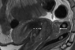
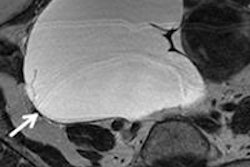
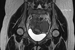
In a new study, Belgian researchers found that MRI can predict the severity of endometriosis in women referred for surgery when it is used as a complement to transvaginal ultrasound. Results were published online on 10 September in Ultrasound in Obstetrics & Gynecology.
The modality works just as well as standard presurgical imaging with intravenous urography (IVU) and double-contrast barium enema (DCBE), but it avoids radiation exposure or the need for contrast, wrote a team led by Dr. Didier Bielen, PhD, of KU Leuven University Hospitals.
" 'One-stop' MRI plus an expert transvaginal ultrasound predicts the intraoperative findings equally well as compared to the standard radiological imaging plus transvaginal ultrasound for patients referred for endometriosis surgery ... without the need of ionizing radiation or intravenous contrast," the group wrote.
In cases of suspected deep endometriosis, European Society of Human Reproduction and Embryology guidelines recommend transvaginal ultrasound plus additional imaging to assess involvement in the ureter, bladder, bowel, and abdominal organs, the authors wrote. This additional imaging tends to be intravenous urography to evaluate the ureter and a double-contrast barium enema to evaluate the rectum, sigmoid, and cecum.
But IVU exposes women to radiation and DCBE requires the administration of contrast. That's why MRI is an intriguing option for preoperative assessment of endometriosis, the group noted.
"Given the promising results of MRI in staging endometriosis, our study evaluated MRI as a noninvasive and nonionizing complement to transvaginal ultrasound for the preoperative staging," the team wrote.
Bielen and colleagues' study included 74 patients with clinically suspected endometriosis with urological and intestinal involvement who were referred for surgery between 2014 and 2015. All of the women underwent transvaginal ultrasound, DCBE, IVU, and abdominal MRI; gynecologists performed the ultrasound exams, while radiologists performed the DCBE, IVU, and MRI exams.
The combination of transvaginal ultrasound and MRI was correct in 86.5% of patients for assessing involvement of the bowel wall, gynecological structures, bladder, and ureters, the researchers found. The combination of transvaginal ultrasound, IVU, and DCBE was correct in 67.6% of patients (p = 0.02).
The group also found the following:
- IVU and DCBE, as well as the combined findings of transvaginal ultrasound and MRI, enabled a "correct stratification for a monodisciplinary approach by gynecologists or multidisciplinary approach in 90.5% of the patients."
- Transvaginal ultrasound and DCBE underestimated the severity of rectal involvement in 2.7% of patients; it overestimated it in 6.8% of patients.
Using MRI with transvaginal ultrasound offers significant benefits, according to the researchers.
"One of the major advantages of this approach is the radiation benefit, given the total lack of ionizing radiation in MRI, with is in contrast to the IVU and DCBE," they concluded. "Second, regarding at least the noncontrast part of the MR examination, the MRI does not rely on sufficient renal function, as opposed to the IVU. It also overcomes the inconveniences of the DCBE, which is rather cumbersome for both the patient and the radiologist."

![Examples of ultrasound findings and techniques. (A) Images in a 39-year-old male patient with a mass in the left thigh. The mass is heterogeneous on the B-mode US image (compared with the patient in D) and showed increased microvascularity (superb microvascular imaging [SMI]) and shear-wave elastography (SWE) values. Undifferentiated pleomorphic sarcoma was diagnosed at biopsy (with pleomorphic rhabdomyosarcoma in surgical specimen). (B) Images in an 18-year-old male patient with a mass in the left leg. The mass is hypoechoic on the B-mode image, with no other findings suggestive of malignancy. The lesion is in contact with the cortex of the tibia, which is slightly irregular. CT revealed a doubtful anteromedial tibial erosion. The microvascular study demonstrated high vascularization, suggestive of malignancy. Periosteal Ewing sarcoma was diagnosed with both histologic and immunohistochemical confirmation. (C) Images in a 69-year-old female patient with a lump growing on the outside of the left leg. Multiple SWE examinations were performed (please note the high values obtained in the measurements, whereas the color map highlights the stiffness relative to adjacent tissues). SMI showed areas of increased vascularization to target for sampling. Undifferentiated spindle cell sarcoma was diagnosed at biopsy, with residual leiomyosarcoma in the surgical specimen after neoadjuvant therapy. (D) Images in a 56-year-old female patient with a mass in the right thigh. The mass is heterogeneous at both B-mode ultrasound (similar to patient A) and MRI (coronal T2-weighted spectral attenuated inversion recovery [SPAIR]; T1-weighted pre-contrast and postcontrast imaging), which even shows uptake after the administration of paramagnetic contrast material, which is traditionally suggestive of malignancy. Low values at SMI and elastography are suggestive of benignity. Spindle cell lipoma was diagnosed at biopsy, with atypical spindle cell lipomatous tumor in the surgical specimen.](https://img.auntminnieeurope.com/mindful/smg/workspaces/default/uploads/2026/08/images-radiol250278fig2.APCFLSvX6p.jpg?auto=format%2Ccompress&fit=crop&h=100&q=70&w=100)




![Examples of ultrasound findings and techniques. (A) Images in a 39-year-old male patient with a mass in the left thigh. The mass is heterogeneous on the B-mode US image (compared with the patient in D) and showed increased microvascularity (superb microvascular imaging [SMI]) and shear-wave elastography (SWE) values. Undifferentiated pleomorphic sarcoma was diagnosed at biopsy (with pleomorphic rhabdomyosarcoma in surgical specimen). (B) Images in an 18-year-old male patient with a mass in the left leg. The mass is hypoechoic on the B-mode image, with no other findings suggestive of malignancy. The lesion is in contact with the cortex of the tibia, which is slightly irregular. CT revealed a doubtful anteromedial tibial erosion. The microvascular study demonstrated high vascularization, suggestive of malignancy. Periosteal Ewing sarcoma was diagnosed with both histologic and immunohistochemical confirmation. (C) Images in a 69-year-old female patient with a lump growing on the outside of the left leg. Multiple SWE examinations were performed (please note the high values obtained in the measurements, whereas the color map highlights the stiffness relative to adjacent tissues). SMI showed areas of increased vascularization to target for sampling. Undifferentiated spindle cell sarcoma was diagnosed at biopsy, with residual leiomyosarcoma in the surgical specimen after neoadjuvant therapy. (D) Images in a 56-year-old female patient with a mass in the right thigh. The mass is heterogeneous at both B-mode ultrasound (similar to patient A) and MRI (coronal T2-weighted spectral attenuated inversion recovery [SPAIR]; T1-weighted pre-contrast and postcontrast imaging), which even shows uptake after the administration of paramagnetic contrast material, which is traditionally suggestive of malignancy. Low values at SMI and elastography are suggestive of benignity. Spindle cell lipoma was diagnosed at biopsy, with atypical spindle cell lipomatous tumor in the surgical specimen.](https://img.auntminnieeurope.com/mindful/smg/workspaces/default/uploads/2026/08/images-radiol250278fig2.APCFLSvX6p.jpg?auto=format%2Ccompress&dpr=2&fit=crop&h=167&q=70&w=250)


