Researchers from Belgium have used the highly photorealistic visualization provided by cinematic rendering technology to enhance their understanding of CT angiography (CTA) scans during postmortem analysis, according to an article recently published online in Diagnostic and Interventional Imaging.
In the case study, clinicians from Université Catholique de Louvain in Brussels acquired the multiphase CTA scans of a 71-year-old woman who died of unknown causes. They decided to use the postmortem CTA data to examine the corpse, as opposed to conducting a conventional autopsy.
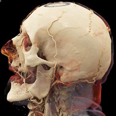
 Cinematically rendered head CT angiography scan of a deceased patient. All images courtesy of Dr. Emmanuel Coche, PhD.
The researchers initially used maximum intensity projection (MIP) to enhance their view of the CTA scans, but they found that the 3D data offered limited visibility of overlapping blood vessels. As a result, they turned to cinematic rendering, a relatively new form of 3D visualization that uses a reconstruction algorithm to propagate light through images from numerous directions (Diagn Interv Imaging, 10 June 2019).
"In the context of forensic medicine optimization in our radiology department, we have naturally thought of applying the cinematic rendering technique to this specific area," senior author Dr. Emmanuel Coche, PhD, told AuntMinnieEurope.com. "We have found the results to be spectacular, with a natural appearance of the different organs and vessels of the deceased patient."
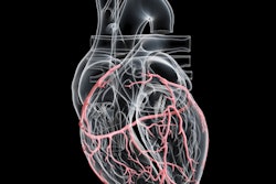
 Cinematically rendered chest CT angiography scan.
After reconstructing the CTA scans using cinematic rendering software (syngo.via VB20, Siemens Healthineers), Coche and colleagues found that the newly reconstructed images offered additional information about the depth of tissues and the state of blood vessels -- especially around the intestines -- previously unrecognized on standard CTA scans.
The cinematically rendered CTA scans delivered a natural appearance and level of transparency that facilitated the comprehension of different anatomical structures, with particularly strong analysis of thoracic organs and vessels, Coche said.
"The major advantage of the cinematic rendering technique is that it delivers a natural appearance of the different tissues with a 3D vision," he said. "The delineation of the vessels is optimal and the relationship with surrounding organs is excellent."
These advantages of cinematic rendering have encouraged the group to begin applying the technique to the analysis of postmortem fetal CTA scans.
Furthermore, producing the cinematically rendered images required a multidisciplinary effort involving the work of radiologic technologists, forensic pathologists, and radiologists, with radiologists primarily serving as image consultants, Coche noted. Optimization of the technique depends on continued discussions among members of the diverse team, promoting collaboration.