U.K. researchers have found that digital breast tomosynthesis (DBT) reduces the benign biopsy rate by as much as 23 percentage points compared with the full-field digital mammography (FFDM) rate, according to a study published online on 19 March in Radiology.
Lower benign biopsy rates would minimize what some call the "harms" of mammography, specifically false positives, lead author Dr. Nisha Sharma of the U.K.'s Leeds Teaching Hospital National Health Service (NHS) Trust told AuntMinnieEurope.com.
"DBT as a screening modality will help to reduce false-positive recalls, and this in turn will minimize harm," Sharma said. "It's an important tool in the diagnostic workup of mammographic lesions, as it provides radiologists with additional information that can reduce the need for biopsy in a significant number of women."
Reducing false positives
The U.K.'s National Health Service Breast Screening Programme (NHSBSP) calls women between the ages of 50 and 70 for screening every three years. DBT is not used in the initial screening phase, although it may be used in follow-up assessment on abnormal mammography, which includes what the NHS calls "triple assessment" -- clinical examination, further imaging, and potential biopsy -- according to Sharma's group.
How DBT affects the benign biopsy rate hasn't been clear, which is why the investigators conducted the study, the group wrote.
"Several studies have compared DBT with FFDM," the team noted. "These studies focused on the need for additional views when using DBT and the ability of DBT to increase the radiologist's confidence in determining if abnormalities are benign or malignant. There is very little published material on the effect of DBT in reducing the benign biopsy rate."
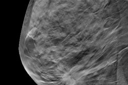
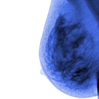
The study included data from 30,933 women who underwent FFDM or screening breast MRI through the NHSBSP at Seacroft Hospital in Leeds between 2015 and 2016. Of these, 1,470 were recalled, for a rate of 4.8%; these women also underwent DBT exams as part of their follow-up evaluation.
Of the 1,470 women recalled, 827 met study inclusion criteria. The cancer detection rate was 17.2% (142 of 827), while the biopsy rate was 69% (571 biopsies in 827 women). In 429 of the biopsies performed, the suspicious lesion detected on screening mammography was not cancerous, for a benign biopsy rate of 75%.
Sharma's team read the DBT images blinded to the original screening results to evaluate whether 3D breast imaging would have influenced the recommendation for biopsy. The group found that DBT would have reduced the number of biopsies performed on recalled women from 571 to 298, without missing the 142 cancers, for a biopsy rate of 36% and a benign biopsy rate of 52%.
| Digital mammography vs. DBT for breast screening biopsy | ||
| Performance measure | FFDM | DBT |
| Recalls | 827 | 245 |
| Biopsies performed | 571 | 298 |
| Screen-detected cancers | 142 | 142 |
| Benign biopsy rate | 75% | 52% |
| Positive predictive value (PPV) of performed biopsies | 24.9% | 47.7% |
| Negative predictive value (NPV)* | 97.9% | 97.9% |
| Sensitivity* | 98.9% | 99.5% |
| Specificity | 38.2% | 77.5% |
"Our results indicate 273 biopsies could have been avoided if DBT had been used," Sharma and colleagues wrote. "This suggests that implementing DBT along with triple assessment could potentially reduce the number of false-positive findings for those abnormalities."
More good news for DBT ... and for women
The study findings demonstrate yet another of DBT's benefits, the researchers concluded.
"DBT has an important role in screening assessment," they wrote. "The key benefit is the reduction in biopsy rate ... without any reduction in the cancer detection rate."