
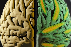
With the help of MRI, researchers from King's College London found that one dose of the cannabis extract cannabidiol may help reduce abnormal brain function in people with psychosis, according to a study published online on August 29 in JAMA Psychiatry.
The researchers analyzed a group of 33 young people who were experiencing distressing psychotic symptoms but had not yet been diagnosed with psychosis. Sixteen subjects received a single dose of cannabidiol, while the other 17 received a placebo. For comparison, the study also included 19 healthy control subjects.
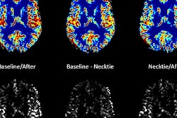
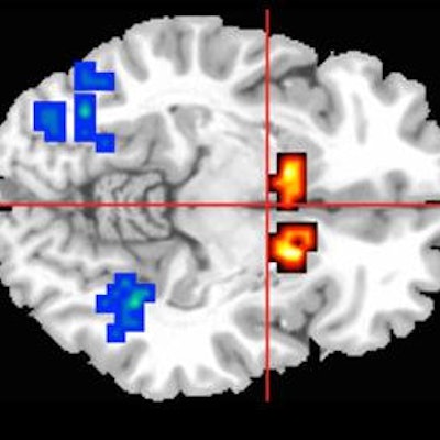
MRI shows activity (red and yellow areas) in the caudate region of the brain, which is affected in people with psychosis. Image courtesy of King's College London.All participants underwent MRI scans while performing a memory task to engage three regions of the brain involved in psychosis.
Among those at risk for psychosis, the subjects who were given cannabidiol showed less-severe abnormal brain activity than those who received a placebo, suggesting that cannabidiol can help readjust brain activity to normal levels.
The researchers are now launching a large-scale, multicenter trial to determine whether cannabidiol can be beneficial in treating young people at high risk of developing psychosis.


![Overview of the study design. (A) The fully automated deep learning framework was developed to estimate body composition (BC) (defined as subcutaneous adipose tissue [SAT] in liters; visceral adipose tissue [VAT] in liters; skeletal muscle [SM] in liters; SM fat fraction [SMFF] as a percentage; and intramuscular adipose tissue [IMAT] in deciliters) from MRI. The fully automated framework comprised one model (model 1) to quantify different BC measures (SAT, VAT, SM, SMFF, and IMAT) as three-dimensional (3D) measures from whole-body MRI scans. The second model (model 2) was trained to identify standardized anatomic landmarks along the craniocaudal body axis (z coordinate field), which allowed for subdividing the whole-body measures into different subregions typically examined on clinical routine MRI scans (chest, abdomen, and pelvis). (B) BC was quantified from whole-body MRI in over 66,000 individuals from two large population-based cohort studies, the UK Biobank (UKB) (36,317 individuals) and the German National Cohort (NAKO) (30,291 individuals). Bar graphs show age distribution by sex and cohort. BMI = body mass index. (C) After the performance assessment of the fully automated framework, the change in BC measures, distributions, and profiles across age decades were investigated. Age-, sex-, and height-adjusted body composition reference curves were calculated and made publicly available in a web-based z-score calculator (https://circ-ml.github.io).](https://img.auntminnieeurope.com/mindful/smg/workspaces/default/uploads/2026/05/body-comp.XgAjTfPj1W.jpg?auto=format%2Ccompress&fit=crop&h=112&q=70&w=112)


