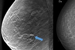
A new meta-analysis has reaffirmed the value of digital breast tomosynthesis (DBT) for breast screening, with DBT detecting more cancers with fewer recalls. But DBT's effect varied significantly between the U.S. and Europe, according to results published online August 9 in the Journal of the National Cancer Institute.
DBT improves cancer detection rates and reduces recalls compared with 2D mammography, but these effects are more pronounced in particular screening settings, wrote a team led by M. Luke Marinovich, PhD, from the University of Sydney. The cancer detection rate (CDR) with DBT was higher in studies from Europe, perhaps due to the longer intervals between screens in Europe, which meant that tomosynthesis caught more cancers.
"The higher incremental CDR attributed to tomosynthesis from the European/Scandinavian studies reflects that, given the longer time interval between screenings, there is greater gain in CDR from tomosynthesis screening," the researchers wrote.
Marinovich's group searched electronic databases between the years 2009 and 2017 for studies that compared tomosynthesis and 2D mammography in asymptomatic women who presented for breast cancer screening. The team identified 17 studies that also reported cancer detection and recall rates for the two modalities, representing 1 million participants.
Overall, tomosynthesis found more cancers than 2D mammography, but the cancer detection rate was higher in Europe than in the U.S. The European studies also tended to use DBT with 2D mammography in the experimental arm, compared with DBT alone in the U.S.
| Cancer detection rate per 1,000 screens of 2D mammography vs. DBT | ||
| Region | 2D mammography | Tomosynthesis |
| European/Scandinavian studies | 6.4 | 8.8 |
| U.S. studies | 4.5 | 5.7 |
As for the recall rate, tomosynthesis led to a reduction in the U.S. studies, but it had little effect in the European/Scandinavian trials.
| Recall rate of 2D mammography vs. DBT | ||
| Region | 2D mammography | Tomosynthesis |
| European/Scandinavian studies | 3.5% | 4.1% |
| U.S. studies | 11.3% | 8% |
The findings suggest that tomosynthesis screening best reduces recall in settings where standard 2D mammography recall rates are relatively high -- as in the U.S. It has a limited effect on recall in screening programs with relatively low recall rates, as in Europe, Marinovich and colleagues wrote.
Although DBT shows clear clinical benefits, more research is needed, according to the investigators.
"Our meta-analysis provides up-to-date pooled estimates of effect for breast cancer screening using tomosynthesis compared with 2D mammography to inform screening research and practice. ... New research in tomosynthesis screening should focus on evaluating both the sustained effect (of repeat screening with tomosynthesis) and the intermediate to long-term outcomes," the group wrote.


![Examples of ultrasound findings and techniques. (A) Images in a 39-year-old male patient with a mass in the left thigh. The mass is heterogeneous on the B-mode US image (compared with the patient in D) and showed increased microvascularity (superb microvascular imaging [SMI]) and shear-wave elastography (SWE) values. Undifferentiated pleomorphic sarcoma was diagnosed at biopsy (with pleomorphic rhabdomyosarcoma in surgical specimen). (B) Images in an 18-year-old male patient with a mass in the left leg. The mass is hypoechoic on the B-mode image, with no other findings suggestive of malignancy. The lesion is in contact with the cortex of the tibia, which is slightly irregular. CT revealed a doubtful anteromedial tibial erosion. The microvascular study demonstrated high vascularization, suggestive of malignancy. Periosteal Ewing sarcoma was diagnosed with both histologic and immunohistochemical confirmation. (C) Images in a 69-year-old female patient with a lump growing on the outside of the left leg. Multiple SWE examinations were performed (please note the high values obtained in the measurements, whereas the color map highlights the stiffness relative to adjacent tissues). SMI showed areas of increased vascularization to target for sampling. Undifferentiated spindle cell sarcoma was diagnosed at biopsy, with residual leiomyosarcoma in the surgical specimen after neoadjuvant therapy. (D) Images in a 56-year-old female patient with a mass in the right thigh. The mass is heterogeneous at both B-mode ultrasound (similar to patient A) and MRI (coronal T2-weighted spectral attenuated inversion recovery [SPAIR]; T1-weighted pre-contrast and postcontrast imaging), which even shows uptake after the administration of paramagnetic contrast material, which is traditionally suggestive of malignancy. Low values at SMI and elastography are suggestive of benignity. Spindle cell lipoma was diagnosed at biopsy, with atypical spindle cell lipomatous tumor in the surgical specimen.](https://img.auntminnieeurope.com/mindful/smg/workspaces/default/uploads/2026/08/images-radiol250278fig2.APCFLSvX6p.jpg?auto=format%2Ccompress&fit=crop&h=100&q=70&w=100)


![Examples of ultrasound findings and techniques. (A) Images in a 39-year-old male patient with a mass in the left thigh. The mass is heterogeneous on the B-mode US image (compared with the patient in D) and showed increased microvascularity (superb microvascular imaging [SMI]) and shear-wave elastography (SWE) values. Undifferentiated pleomorphic sarcoma was diagnosed at biopsy (with pleomorphic rhabdomyosarcoma in surgical specimen). (B) Images in an 18-year-old male patient with a mass in the left leg. The mass is hypoechoic on the B-mode image, with no other findings suggestive of malignancy. The lesion is in contact with the cortex of the tibia, which is slightly irregular. CT revealed a doubtful anteromedial tibial erosion. The microvascular study demonstrated high vascularization, suggestive of malignancy. Periosteal Ewing sarcoma was diagnosed with both histologic and immunohistochemical confirmation. (C) Images in a 69-year-old female patient with a lump growing on the outside of the left leg. Multiple SWE examinations were performed (please note the high values obtained in the measurements, whereas the color map highlights the stiffness relative to adjacent tissues). SMI showed areas of increased vascularization to target for sampling. Undifferentiated spindle cell sarcoma was diagnosed at biopsy, with residual leiomyosarcoma in the surgical specimen after neoadjuvant therapy. (D) Images in a 56-year-old female patient with a mass in the right thigh. The mass is heterogeneous at both B-mode ultrasound (similar to patient A) and MRI (coronal T2-weighted spectral attenuated inversion recovery [SPAIR]; T1-weighted pre-contrast and postcontrast imaging), which even shows uptake after the administration of paramagnetic contrast material, which is traditionally suggestive of malignancy. Low values at SMI and elastography are suggestive of benignity. Spindle cell lipoma was diagnosed at biopsy, with atypical spindle cell lipomatous tumor in the surgical specimen.](https://img.auntminnieeurope.com/mindful/smg/workspaces/default/uploads/2026/08/images-radiol250278fig2.APCFLSvX6p.jpg?auto=format%2Ccompress&fit=crop&h=112&q=70&w=112)