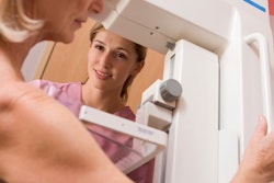
Breast cancer screening attendance in Switzerland has hit a new low, despite expanding regional mammography screening programs. Several breast radiologists have blamed antiscreening information and the debate surrounding the pros and cons of breast cancer screening.
Swiss researchers analyzed data from nationwide representative health surveys. Led by Dr. Lukas Fenner, a senior research scientist at the Institute of Social and Preventive Medicine, University of Bern, the researchers spotted a drop of more than 7% in mammography use from 2007 to 2012.
"This decline might reflect the ongoing debate about the benefits (mortality) and harms (false-positive results requiring diagnostic workup, overdiagnosis, overtreatment) of breast cancer screening, and the increasing emphasis that is put on ensuring informed decision-making," he and his colleagues wrote (Swiss Medical Weekly, 14 May 2018).
Regional screening
In Switzerland, screening attendance is higher in the French-speaking area, which has screening programs, than in the German-speaking area, which has mainly opportunistic screening, and this difference does not appear to be explained by sociodemographic or health-related factors, according to Fenner.
Swiss national strategy reports have repeatedly suggested the adoption of a national screening program to no avail. Swiss cantons, similar to counties, may or may not implement a program independently. Breast cancer screening attendance was expected to change after cantonal mammography programs were expanded, slowly growing to 11 out of 26 cantons as of 2012.
Fenner and colleagues studied time trends and the associations of sociodemographic characteristics and health-related factors with the changes in breast cancer screening in Switzerland between 2007 and 2012, using data from the nationwide representative health surveys.
They included 11,292 female respondents ages 40 to 79 who responded to a written questionnaire. For each survey year, 2007 and 2012, they calculated the proportions of the female population who reported a mammogram in the past 12 months, the proportion of mammography as part of a screening program, and the proportion of screening mammography initiated by the woman or a physician. They used univariate and multivariate logistic regression models to calculate unadjusted and adjusted odds ratios.
The researchers found that the use of any mammography in the past 12 months declined from 19.1% in 2007 to 11.7% in 2012. The decline was more pronounced in regions with a long-standing program or no cantonal breast cancer screening program; levels remained relatively stable in regions with a recently introduced program.
As expected, the regions with long-standing screening programs corresponded to the French-speaking area of the country and had a high uptake, whereas opportunistic screening dominated in the German-speaking area, Fenner and colleagues noted.
"This difference did not seem to be influenced by sociodemographic or health-related factors, but appears to be driven by public promotion of screening, recommendations by healthcare providers, and cultural factors, including different beliefs and attitudes," they wrote.
The researchers also found that opportunistic screening dropped from 12.0% in 2007 to 6.2% in 2012, whereas the use of programmatic mammography remained stable at 3.1%. Use of any mammography was higher in women ages 50 to 69, those residing in a region with a systematic screening program in place, and those having a private hospital stay insurance, but it was not associated with education level and non-Swiss citizenship.
The results suggest at least a temporary positive effect of the established cantonal screening programs, particularly in the more recently introduced programs, but also a shift from individual screening to cantonal programs, according to the researchers.
They also noted that the level of information given to the public appears to strongly influence the willingness to participate in screening programs.
"The positive effects of breast cancer screening were initially overestimated by women in Switzerland and elsewhere, but this may have changed over time to a more critical public view on breast cancer screening," they wrote.
An example of a critical view of breast cancer screening comes from the Swiss Medical Board, which in 2014 recommended winding down breast cancer screening programs. That announcement came after data were collected for the current study.
Observers give views
The results correspond with the perspective of several breast radiologists.
 Dr. Gábor Forrai, PhD, president of the EUSOBI.
"I am not surprised by the negative reflection of the women in general," said Dr. Gábor Forrai, PhD, president of the European Society of Breast Imaging (EUSOBI).
He said information does not exist in a vacuum and pointed to what happened when actress Angelina Jolie found out she was at high risk for breast cancer: Screening attendance increased, and at his center, the scheduling time almost doubled.
"We can easily suppose the negative public information leads to the same effect: significant decrease of attendance," he said. "That's why it is a very sensitive subject. What these 'sensation-hunting' persons and speakers do not feel: If they campaign against screening, they withdraw a certain number of women from attending it -- therefore all these women's breast cancers remain undiscovered. So the potential loss of their life remains these irresponsible people's responsibility."
Speaking more generally about Switzerland, Dr. Karen Kinkel, PhD, from the radiology institute at Clinique des Grangettes in Chêne-Bougeries, Switzerland, said the canton of Geneva has good quality individual breast screening. However, "In the German-speaking part of Switzerland, politicians were very much influenced by antiscreening publications from Germany."
In terms of future research, Fenner and colleagues noted that data from the next survey round of 2017 will be available at the end of this year and might show further trends. Future surveys should include additional questions to better distinguish between diagnostic, opportunistic, and programmatic mammography, as well as questions on the willingness to participate in screening programs.