Dear AuntMinnieEurope Member,
A lengthy article about the safety of gadolinium-based contrast agents appeared in a German newspaper last week. The headline referred to "a poisoned atmosphere in the (MRI) tube," and it followed an earlier article that boldly asserted "an MRI shot can be poisonous."
It's tempting to dismiss such material as sensationalism, but the important point is that many patients are reading these reports. Radiologists and radiographers must know what's being written about gadolinium and be prepared for the awkward questions. To read the Maverinck's new column, go to the MRI Community, or click here.
Another MRI-related topic being followed closely by patients is the death of a man pulled into the bore of a scanner in an Indian hospital. Click here for the latest on this story.
Meanwhile, a large U.K. trial has shown that extended ipsilateral breast ultrasound can detect many extra cancers compared with the standard screening protocol, but less effect was demonstrated for complete contralateral breast ultrasound. Visit the Women's Imaging Community, or click here.
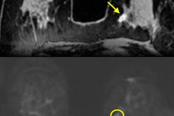
Breast MRI has a high sensitivity, whether that's dynamic contrast-enhanced MRI (DCE-MRI) or diffusion-weighted imaging (DWI-MRI), but a drawback is that DCE-MRI tends to pick up everything -- benign or malignant. DWI-MRI doesn't detect everything, but it accurately distinguishes between benign and malignant breast lesions. Researchers from the U.S. and Austria have looked at what happens when these two techniques are put together. Get the full details here.
Hyperechogenic breast lesions are rare and described as generally benign; these lesions are often overlooked and a biopsy is not generally recommended. New research suggests it is not the ideal approach. Investigators found that hyperechogenic lesions can be malignant and must be imaged with that possibility in mind.
Finally, don't miss our case report about a 71-year-old woman found unconscious in her bed. This original case came from Dr. Fatih Seker in Heidelberg, Germany, who is a member of our editorial advisory board. Click here to test yourself.


![Examples of ultrasound findings and techniques. (A) Images in a 39-year-old male patient with a mass in the left thigh. The mass is heterogeneous on the B-mode US image (compared with the patient in D) and showed increased microvascularity (superb microvascular imaging [SMI]) and shear-wave elastography (SWE) values. Undifferentiated pleomorphic sarcoma was diagnosed at biopsy (with pleomorphic rhabdomyosarcoma in surgical specimen). (B) Images in an 18-year-old male patient with a mass in the left leg. The mass is hypoechoic on the B-mode image, with no other findings suggestive of malignancy. The lesion is in contact with the cortex of the tibia, which is slightly irregular. CT revealed a doubtful anteromedial tibial erosion. The microvascular study demonstrated high vascularization, suggestive of malignancy. Periosteal Ewing sarcoma was diagnosed with both histologic and immunohistochemical confirmation. (C) Images in a 69-year-old female patient with a lump growing on the outside of the left leg. Multiple SWE examinations were performed (please note the high values obtained in the measurements, whereas the color map highlights the stiffness relative to adjacent tissues). SMI showed areas of increased vascularization to target for sampling. Undifferentiated spindle cell sarcoma was diagnosed at biopsy, with residual leiomyosarcoma in the surgical specimen after neoadjuvant therapy. (D) Images in a 56-year-old female patient with a mass in the right thigh. The mass is heterogeneous at both B-mode ultrasound (similar to patient A) and MRI (coronal T2-weighted spectral attenuated inversion recovery [SPAIR]; T1-weighted pre-contrast and postcontrast imaging), which even shows uptake after the administration of paramagnetic contrast material, which is traditionally suggestive of malignancy. Low values at SMI and elastography are suggestive of benignity. Spindle cell lipoma was diagnosed at biopsy, with atypical spindle cell lipomatous tumor in the surgical specimen.](https://img.auntminnieeurope.com/mindful/smg/workspaces/default/uploads/2026/08/images-radiol250278fig2.APCFLSvX6p.jpg?auto=format%2Ccompress&fit=crop&h=100&q=70&w=100)


![Examples of ultrasound findings and techniques. (A) Images in a 39-year-old male patient with a mass in the left thigh. The mass is heterogeneous on the B-mode US image (compared with the patient in D) and showed increased microvascularity (superb microvascular imaging [SMI]) and shear-wave elastography (SWE) values. Undifferentiated pleomorphic sarcoma was diagnosed at biopsy (with pleomorphic rhabdomyosarcoma in surgical specimen). (B) Images in an 18-year-old male patient with a mass in the left leg. The mass is hypoechoic on the B-mode image, with no other findings suggestive of malignancy. The lesion is in contact with the cortex of the tibia, which is slightly irregular. CT revealed a doubtful anteromedial tibial erosion. The microvascular study demonstrated high vascularization, suggestive of malignancy. Periosteal Ewing sarcoma was diagnosed with both histologic and immunohistochemical confirmation. (C) Images in a 69-year-old female patient with a lump growing on the outside of the left leg. Multiple SWE examinations were performed (please note the high values obtained in the measurements, whereas the color map highlights the stiffness relative to adjacent tissues). SMI showed areas of increased vascularization to target for sampling. Undifferentiated spindle cell sarcoma was diagnosed at biopsy, with residual leiomyosarcoma in the surgical specimen after neoadjuvant therapy. (D) Images in a 56-year-old female patient with a mass in the right thigh. The mass is heterogeneous at both B-mode ultrasound (similar to patient A) and MRI (coronal T2-weighted spectral attenuated inversion recovery [SPAIR]; T1-weighted pre-contrast and postcontrast imaging), which even shows uptake after the administration of paramagnetic contrast material, which is traditionally suggestive of malignancy. Low values at SMI and elastography are suggestive of benignity. Spindle cell lipoma was diagnosed at biopsy, with atypical spindle cell lipomatous tumor in the surgical specimen.](https://img.auntminnieeurope.com/mindful/smg/workspaces/default/uploads/2026/08/images-radiol250278fig2.APCFLSvX6p.jpg?auto=format%2Ccompress&fit=crop&h=112&q=70&w=112)