Dear AuntMinnieEurope Member,
Soaring temperatures of 30° C were the order of the day when the German Radiology Congress drew to a close at the weekend, and we've posted articles about the following red-hot presentations from the meeting:
- Safety of MRI contrast agents. For details about a new study from Düsseldorf, go to our MRI Community, or click here.
- Use of advanced software for evaluating CT scans of stroke patients to assess stroke severity and help identify patients who will not benefit from thrombectomy. Visit the CT Community, or click here.
- The clinical potential of phase-contrast imaging, as explained by Franz Pfeiffer, PhD, professor of biomedical physics at the Technical University of Munich. Get the details here.
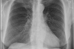
Elsewhere, Dutch researchers have published important findings on the diagnosis of chronic obstructive pulmonary disease (COPD). They think their study has raised awareness at their hospital of the challenges of diagnosing COPD on a chest x-ray, and they urge all radiologists to be wary of mentioning suspicion of COPD in chest radiograph reports. Click here to learn more.
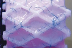
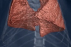
Three-dimensional printing continues to evolve rapidly, and is now being proposed to create cardiac stents for use in children. Another Dutch group has warned that many hurdles still need to be overcome, but they've created a new 3D-printed polymer stent that bypasses many of the limitations of conventional nitinol stents for use with tissue-engineered heart valves. Read more about the project in the Advanced Visualization Community, or click here.
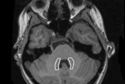
Finally, don't miss our Case of the Week, a middle-aged alcoholic man who was found collapsed at home. Click here to test yourself.


![Overview of the study design. (A) The fully automated deep learning framework was developed to estimate body composition (BC) (defined as subcutaneous adipose tissue [SAT] in liters; visceral adipose tissue [VAT] in liters; skeletal muscle [SM] in liters; SM fat fraction [SMFF] as a percentage; and intramuscular adipose tissue [IMAT] in deciliters) from MRI. The fully automated framework comprised one model (model 1) to quantify different BC measures (SAT, VAT, SM, SMFF, and IMAT) as three-dimensional (3D) measures from whole-body MRI scans. The second model (model 2) was trained to identify standardized anatomic landmarks along the craniocaudal body axis (z coordinate field), which allowed for subdividing the whole-body measures into different subregions typically examined on clinical routine MRI scans (chest, abdomen, and pelvis). (B) BC was quantified from whole-body MRI in over 66,000 individuals from two large population-based cohort studies, the UK Biobank (UKB) (36,317 individuals) and the German National Cohort (NAKO) (30,291 individuals). Bar graphs show age distribution by sex and cohort. BMI = body mass index. (C) After the performance assessment of the fully automated framework, the change in BC measures, distributions, and profiles across age decades were investigated. Age-, sex-, and height-adjusted body composition reference curves were calculated and made publicly available in a web-based z-score calculator (https://circ-ml.github.io).](https://img.auntminnieeurope.com/mindful/smg/workspaces/default/uploads/2026/05/body-comp.XgAjTfPj1W.jpg?auto=format%2Ccompress&fit=crop&h=100&q=70&w=100)


![Overview of the study design. (A) The fully automated deep learning framework was developed to estimate body composition (BC) (defined as subcutaneous adipose tissue [SAT] in liters; visceral adipose tissue [VAT] in liters; skeletal muscle [SM] in liters; SM fat fraction [SMFF] as a percentage; and intramuscular adipose tissue [IMAT] in deciliters) from MRI. The fully automated framework comprised one model (model 1) to quantify different BC measures (SAT, VAT, SM, SMFF, and IMAT) as three-dimensional (3D) measures from whole-body MRI scans. The second model (model 2) was trained to identify standardized anatomic landmarks along the craniocaudal body axis (z coordinate field), which allowed for subdividing the whole-body measures into different subregions typically examined on clinical routine MRI scans (chest, abdomen, and pelvis). (B) BC was quantified from whole-body MRI in over 66,000 individuals from two large population-based cohort studies, the UK Biobank (UKB) (36,317 individuals) and the German National Cohort (NAKO) (30,291 individuals). Bar graphs show age distribution by sex and cohort. BMI = body mass index. (C) After the performance assessment of the fully automated framework, the change in BC measures, distributions, and profiles across age decades were investigated. Age-, sex-, and height-adjusted body composition reference curves were calculated and made publicly available in a web-based z-score calculator (https://circ-ml.github.io).](https://img.auntminnieeurope.com/mindful/smg/workspaces/default/uploads/2026/05/body-comp.XgAjTfPj1W.jpg?auto=format%2Ccompress&fit=crop&h=112&q=70&w=112)


