Dear Cardiac Imaging Insider,
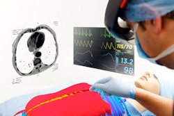
Robust growth in cardiac imaging means increased training needs for radiologists and trainees looking to fill the gap in qualified imaging personnel. Unfortunately, however, training programs are not where they need to be. Trainees are often rushed, with little time to look at cases, and the teachers don't always focus on the most critical knowledge -- like which imaging findings are important enough to warrant a change in management.
But the team at Barts London Chest Hospital in the U.K. seems to have things figured out. And you can find out what they've learned about cardiac imaging by clicking here.
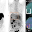
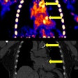
In the Netherlands, researchers used SPECT gamma cameras with cadmium zinc telluride-based (CZT) digital detector technology to confirm that the degree of abnormal myocardial perfusion imaging increased along with chances of an adverse event within three years. What's more, CZT-based SPECT achieved the results with less radiation exposure for cardiac patients and no degradation in diagnostic image quality.
For a broader look at how CZT-based SPECT is heralding new discoveries that will benefit a wide range of patients, click here.
Speaking of SPECT, a large nuclear cardiology lab was able to cut its average dose by more than 60%, using nothing more complicated than common-sense steps available to everyone. One common step was skipping the rest part of a stress-rest exam whenever possible. And there are others, including some that are less well known. Click here for advice on reducing nuclear cardiology doses to patients.
In cardiac CT perfusion imaging, many clinicians have been impressed by results showing excellent contrast properties, but consider CT perfusion to be a relatively high-dose exam suitable only for special needs. But that's no longer true, according to a study presented at ECR 2017. Using the latest-generation dual-source CT scanner, researchers found they could acquire excellent images at surprisingly low radiation doses. Get the rest of the story here.
Another important ECR 2017 presentation discussed key cardiac findings in stroke. Advances in CT and MRI mean it's now possible to identify subtle cardiac pathologies responsible for stroke that once remained unseen. The new discoveries are putting greater emphasis on the ability of imaging professionals to find them, said Spanish researchers who received an award for their work at the Vienna meeting.
In Germany, investigators are using advanced MRI techniques to distinguish acute from chronic myocardial infarction. Find out how they do it by clicking here. Finally, a study from the Netherlands showed how a couple of simple techniques can reduce the need for iodinated contrast across different CT platforms.
And that's just the beginning. We invite you to scroll through the links below for more news from the heart of cardiac imaging in Europe, all here in your Cardiac Imaging Community.

