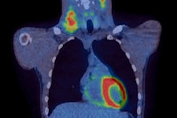
Using coronary CT angiography (CTA), Dutch researchers have found that Hodgkin's lymphoma survivors have more severe coronary artery disease (CAD) 20 years after chest irradiation
The research, presented at the International Conference on Nuclear Cardiology and Cardiac CT (ICNC) 2017, assessed the extent, severity, and location of coronary artery disease in Hodgkin's lymphoma survivors who had received chest irradiation. The study included 79 patients who had been free of Hodgkin's lymphoma for at least 10 years and received mediastinal irradiation 20 years ago, plus 273 controls without Hodgkin's lymphoma or irradiation.
"Patients with Hodgkin's lymphoma receive high-dose mediastinal irradiation at a young age as part of their treatment," said Dr. Alexander van Rosendael, from Leiden University Medical Centre in the Netherlands. "There is an ongoing debate about whether to screen patients who get chest irradiation for coronary artery disease."
Patients and controls were matched in a one-to-three fashion by age, gender, diabetes, hypertension, hypercholesterolemia, family history of coronary artery disease, and smoking status. Patients were 45 years old on average, and the presence of cardiovascular risk factors was low overall. Just 42% of patients had no atherosclerosis on coronary CTA compared with 64% of the controls.
Hodgkin's patients had significantly more multivessel CAD: 10% had two-vessel disease and 24% had three-vessel disease compared with 6% and 9% of the controls, respectively. The segment involvement score and the segment stenosis score were significantly higher for Hodgkin's patients.
Compared with the controls, Hodgkin's patients had significantly more coronary plaques in the left main (17% versus 6%), proximal left anterior descending (30% versus 16%), proximal right coronary artery (25% versus 10%), and proximal left circumflex (14% versus 6%) but not in nonproximal coronary segments. Patients had a fourfold risk of proximal plaque and a threefold risk of proximal obstructive stenosis compared with the controls.
Irradiated patients had all the features of high-risk CAD, including high stenosis severity, proximal location, and extensive disease. This may explain why Hodgkin's patients have such poor cardiovascular outcomes when they get older, according to the study authors.
The finding of more, and more severe, CAD in irradiated patients supports the argument for screening, they noted.


![Examples of ultrasound findings and techniques. (A) Images in a 39-year-old male patient with a mass in the left thigh. The mass is heterogeneous on the B-mode US image (compared with the patient in D) and showed increased microvascularity (superb microvascular imaging [SMI]) and shear-wave elastography (SWE) values. Undifferentiated pleomorphic sarcoma was diagnosed at biopsy (with pleomorphic rhabdomyosarcoma in surgical specimen). (B) Images in an 18-year-old male patient with a mass in the left leg. The mass is hypoechoic on the B-mode image, with no other findings suggestive of malignancy. The lesion is in contact with the cortex of the tibia, which is slightly irregular. CT revealed a doubtful anteromedial tibial erosion. The microvascular study demonstrated high vascularization, suggestive of malignancy. Periosteal Ewing sarcoma was diagnosed with both histologic and immunohistochemical confirmation. (C) Images in a 69-year-old female patient with a lump growing on the outside of the left leg. Multiple SWE examinations were performed (please note the high values obtained in the measurements, whereas the color map highlights the stiffness relative to adjacent tissues). SMI showed areas of increased vascularization to target for sampling. Undifferentiated spindle cell sarcoma was diagnosed at biopsy, with residual leiomyosarcoma in the surgical specimen after neoadjuvant therapy. (D) Images in a 56-year-old female patient with a mass in the right thigh. The mass is heterogeneous at both B-mode ultrasound (similar to patient A) and MRI (coronal T2-weighted spectral attenuated inversion recovery [SPAIR]; T1-weighted pre-contrast and postcontrast imaging), which even shows uptake after the administration of paramagnetic contrast material, which is traditionally suggestive of malignancy. Low values at SMI and elastography are suggestive of benignity. Spindle cell lipoma was diagnosed at biopsy, with atypical spindle cell lipomatous tumor in the surgical specimen.](https://img.auntminnieeurope.com/mindful/smg/workspaces/default/uploads/2026/08/images-radiol250278fig2.APCFLSvX6p.jpg?auto=format%2Ccompress&fit=crop&h=100&q=70&w=100)


![Examples of ultrasound findings and techniques. (A) Images in a 39-year-old male patient with a mass in the left thigh. The mass is heterogeneous on the B-mode US image (compared with the patient in D) and showed increased microvascularity (superb microvascular imaging [SMI]) and shear-wave elastography (SWE) values. Undifferentiated pleomorphic sarcoma was diagnosed at biopsy (with pleomorphic rhabdomyosarcoma in surgical specimen). (B) Images in an 18-year-old male patient with a mass in the left leg. The mass is hypoechoic on the B-mode image, with no other findings suggestive of malignancy. The lesion is in contact with the cortex of the tibia, which is slightly irregular. CT revealed a doubtful anteromedial tibial erosion. The microvascular study demonstrated high vascularization, suggestive of malignancy. Periosteal Ewing sarcoma was diagnosed with both histologic and immunohistochemical confirmation. (C) Images in a 69-year-old female patient with a lump growing on the outside of the left leg. Multiple SWE examinations were performed (please note the high values obtained in the measurements, whereas the color map highlights the stiffness relative to adjacent tissues). SMI showed areas of increased vascularization to target for sampling. Undifferentiated spindle cell sarcoma was diagnosed at biopsy, with residual leiomyosarcoma in the surgical specimen after neoadjuvant therapy. (D) Images in a 56-year-old female patient with a mass in the right thigh. The mass is heterogeneous at both B-mode ultrasound (similar to patient A) and MRI (coronal T2-weighted spectral attenuated inversion recovery [SPAIR]; T1-weighted pre-contrast and postcontrast imaging), which even shows uptake after the administration of paramagnetic contrast material, which is traditionally suggestive of malignancy. Low values at SMI and elastography are suggestive of benignity. Spindle cell lipoma was diagnosed at biopsy, with atypical spindle cell lipomatous tumor in the surgical specimen.](https://img.auntminnieeurope.com/mindful/smg/workspaces/default/uploads/2026/08/images-radiol250278fig2.APCFLSvX6p.jpg?auto=format%2Ccompress&fit=crop&h=112&q=70&w=112)