Dear Molecular Imaging Insider,
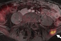
The value of PET/MRI continues to expand in the realm of women's imaging. German researchers have found that restaging breast cancer patients with the hybrid modality is superior to MRI alone. Also, they think the inclusion of MRI sequences such as diffusion-weighted imaging can extend scanning time with debatably beneficial results.
Overall, they endorse PET/MRI for its excellent diagnostic accuracy and diagnostic confidence for the identification of recurrent breast cancer lesions. Read how PET/MRI performs.
PET/MRI, of course, has shown its value in other clinical applications. For example, adding a scan of the entire head to a routine whole-body protocol reveals a significant number of incidental findings in the brains of asymptomatic cancer patients. Researchers recommend expanding a typical whole-body FDG-PET/MRI scan to include the head because the previously unknown findings can have implications for patient treatment or prognosis.
Interestingly, another German team has found that PET/MRI's performance in evaluating head and neck cancer appears no better than that of tried-and-true PET/CT. The group from the University of Düsseldorf found no statistically significant differences between FDG-PET/MRI and FDG-PET/CT in this area.
While PET already is widely used to track molecules within the body with high sensitivity and to image tumors with the help of the radiotracer F-18 FDG, one observer says the modality's use for molecular imaging is just getting started. Charalampos (Harry) Tsoumpas, PhD, a lecturer in medical imaging at the University of Leeds in the U.K., believes PET has much more to offer for other clinical applications as well.
Meanwhile, a technology known as photoacoustic CT can provide noninvasive real-time imaging of significant anatomical and physiological information through strong acoustic waves generated by a pulsed laser. There are, however, disadvantages, such as bulk and cost, but these issues can be solved, experts insist.
Be sure to visit the Molecular Imaging Community on a daily basis to stay informed on the latest news and research from around the continent and the world.


![Examples of ultrasound findings and techniques. (A) Images in a 39-year-old male patient with a mass in the left thigh. The mass is heterogeneous on the B-mode US image (compared with the patient in D) and showed increased microvascularity (superb microvascular imaging [SMI]) and shear-wave elastography (SWE) values. Undifferentiated pleomorphic sarcoma was diagnosed at biopsy (with pleomorphic rhabdomyosarcoma in surgical specimen). (B) Images in an 18-year-old male patient with a mass in the left leg. The mass is hypoechoic on the B-mode image, with no other findings suggestive of malignancy. The lesion is in contact with the cortex of the tibia, which is slightly irregular. CT revealed a doubtful anteromedial tibial erosion. The microvascular study demonstrated high vascularization, suggestive of malignancy. Periosteal Ewing sarcoma was diagnosed with both histologic and immunohistochemical confirmation. (C) Images in a 69-year-old female patient with a lump growing on the outside of the left leg. Multiple SWE examinations were performed (please note the high values obtained in the measurements, whereas the color map highlights the stiffness relative to adjacent tissues). SMI showed areas of increased vascularization to target for sampling. Undifferentiated spindle cell sarcoma was diagnosed at biopsy, with residual leiomyosarcoma in the surgical specimen after neoadjuvant therapy. (D) Images in a 56-year-old female patient with a mass in the right thigh. The mass is heterogeneous at both B-mode ultrasound (similar to patient A) and MRI (coronal T2-weighted spectral attenuated inversion recovery [SPAIR]; T1-weighted pre-contrast and postcontrast imaging), which even shows uptake after the administration of paramagnetic contrast material, which is traditionally suggestive of malignancy. Low values at SMI and elastography are suggestive of benignity. Spindle cell lipoma was diagnosed at biopsy, with atypical spindle cell lipomatous tumor in the surgical specimen.](https://img.auntminnieeurope.com/mindful/smg/workspaces/default/uploads/2026/08/images-radiol250278fig2.APCFLSvX6p.jpg?auto=format%2Ccompress&fit=crop&h=100&q=70&w=100)


![Examples of ultrasound findings and techniques. (A) Images in a 39-year-old male patient with a mass in the left thigh. The mass is heterogeneous on the B-mode US image (compared with the patient in D) and showed increased microvascularity (superb microvascular imaging [SMI]) and shear-wave elastography (SWE) values. Undifferentiated pleomorphic sarcoma was diagnosed at biopsy (with pleomorphic rhabdomyosarcoma in surgical specimen). (B) Images in an 18-year-old male patient with a mass in the left leg. The mass is hypoechoic on the B-mode image, with no other findings suggestive of malignancy. The lesion is in contact with the cortex of the tibia, which is slightly irregular. CT revealed a doubtful anteromedial tibial erosion. The microvascular study demonstrated high vascularization, suggestive of malignancy. Periosteal Ewing sarcoma was diagnosed with both histologic and immunohistochemical confirmation. (C) Images in a 69-year-old female patient with a lump growing on the outside of the left leg. Multiple SWE examinations were performed (please note the high values obtained in the measurements, whereas the color map highlights the stiffness relative to adjacent tissues). SMI showed areas of increased vascularization to target for sampling. Undifferentiated spindle cell sarcoma was diagnosed at biopsy, with residual leiomyosarcoma in the surgical specimen after neoadjuvant therapy. (D) Images in a 56-year-old female patient with a mass in the right thigh. The mass is heterogeneous at both B-mode ultrasound (similar to patient A) and MRI (coronal T2-weighted spectral attenuated inversion recovery [SPAIR]; T1-weighted pre-contrast and postcontrast imaging), which even shows uptake after the administration of paramagnetic contrast material, which is traditionally suggestive of malignancy. Low values at SMI and elastography are suggestive of benignity. Spindle cell lipoma was diagnosed at biopsy, with atypical spindle cell lipomatous tumor in the surgical specimen.](https://img.auntminnieeurope.com/mindful/smg/workspaces/default/uploads/2026/08/images-radiol250278fig2.APCFLSvX6p.jpg?auto=format%2Ccompress&fit=crop&h=112&q=70&w=112)