Dear Advanced Visualization Insider,
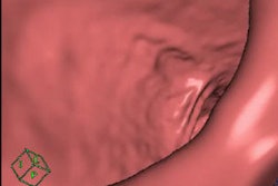
Electronic cleansing has been something of a holy grail in CT colonography (CTC) research for years -- mainly because perfecting the process would allow most CTC exams to be performed without cathartic bowel cleansing, making it easier on patients and attracting people who have been avoiding screening altogether.
Electronic cleansing algorithms work by virtually removing residual matter from colonic CT images, allowing radiologists to see the polyps hidden underneath. Unfortunately, they don't work perfectly.
Now researchers from Italy, Japan, and the U.S. have teamed up to find an algorithm to make electronic cleansing work. Their technique uses deep convolutional neural networks to separate colonic contents. Learn how it performed in a study by clicking here.
A machine-learning technique also boosted the performance of cardiac fractional flow-reserve CT software better than two conventional methods that already work pretty well, according to another study. On a per-patient basis, the technique was superior in sensitivity, specificity, and positive and negative predictive values compared with traditional algorithms. Machine-learning algorithms appear to be skilled at learning patterns in medical images that are characteristic of disease. Maybe that's why they appear to be turning up everywhere.
In body PET/MRI scans, adding the head can reveal a wide array of new incidental findings, from vascular disease to microvascular ischemic changes and brain infarcts, according to researchers. And it hardly adds any time to the body scan.
Three-dimensional printing is opening new possibilities in surgery planning with its ability to enhance the understanding of complex pathologies for referring physicians, residents, students, and patients. But you have to work hard to get it right, said researchers from Switzerland who discuss the challenges of turning a volumetric dataset into a 3D-printed model and determining who will benefit most from the labor-intensive practice.
In Germany, a low-cost 3D printing phantom is being used with quantitative SPECT/CT to optimize radiation dose for radionuclide therapies for patients of all sizes.
In Italy, researchers have developed a prostate cancer computer-aided detection scheme that can improve the chances of selecting the right biopsy targets. The improvement wasn't seen among all readers and all lesions, however. Find out more here.
Why was so much of the recent RSNA 2016 meeting devoted to advanced visualization software? There are many reasons, of course, but in an interoperable world where clinicians can be anywhere, accessibility may be the most important reason. Learn more in contributing writer Dr. Stephen Holloway's article here.
For the rest of the news in 3D, advanced visualization, and computer-aided detection, we invite you to scroll thorough the links below, right here in your Advanced Visualization Community.


![Examples of ultrasound findings and techniques. (A) Images in a 39-year-old male patient with a mass in the left thigh. The mass is heterogeneous on the B-mode US image (compared with the patient in D) and showed increased microvascularity (superb microvascular imaging [SMI]) and shear-wave elastography (SWE) values. Undifferentiated pleomorphic sarcoma was diagnosed at biopsy (with pleomorphic rhabdomyosarcoma in surgical specimen). (B) Images in an 18-year-old male patient with a mass in the left leg. The mass is hypoechoic on the B-mode image, with no other findings suggestive of malignancy. The lesion is in contact with the cortex of the tibia, which is slightly irregular. CT revealed a doubtful anteromedial tibial erosion. The microvascular study demonstrated high vascularization, suggestive of malignancy. Periosteal Ewing sarcoma was diagnosed with both histologic and immunohistochemical confirmation. (C) Images in a 69-year-old female patient with a lump growing on the outside of the left leg. Multiple SWE examinations were performed (please note the high values obtained in the measurements, whereas the color map highlights the stiffness relative to adjacent tissues). SMI showed areas of increased vascularization to target for sampling. Undifferentiated spindle cell sarcoma was diagnosed at biopsy, with residual leiomyosarcoma in the surgical specimen after neoadjuvant therapy. (D) Images in a 56-year-old female patient with a mass in the right thigh. The mass is heterogeneous at both B-mode ultrasound (similar to patient A) and MRI (coronal T2-weighted spectral attenuated inversion recovery [SPAIR]; T1-weighted pre-contrast and postcontrast imaging), which even shows uptake after the administration of paramagnetic contrast material, which is traditionally suggestive of malignancy. Low values at SMI and elastography are suggestive of benignity. Spindle cell lipoma was diagnosed at biopsy, with atypical spindle cell lipomatous tumor in the surgical specimen.](https://img.auntminnieeurope.com/mindful/smg/workspaces/default/uploads/2026/08/images-radiol250278fig2.APCFLSvX6p.jpg?auto=format%2Ccompress&fit=crop&h=100&q=70&w=100)


![Examples of ultrasound findings and techniques. (A) Images in a 39-year-old male patient with a mass in the left thigh. The mass is heterogeneous on the B-mode US image (compared with the patient in D) and showed increased microvascularity (superb microvascular imaging [SMI]) and shear-wave elastography (SWE) values. Undifferentiated pleomorphic sarcoma was diagnosed at biopsy (with pleomorphic rhabdomyosarcoma in surgical specimen). (B) Images in an 18-year-old male patient with a mass in the left leg. The mass is hypoechoic on the B-mode image, with no other findings suggestive of malignancy. The lesion is in contact with the cortex of the tibia, which is slightly irregular. CT revealed a doubtful anteromedial tibial erosion. The microvascular study demonstrated high vascularization, suggestive of malignancy. Periosteal Ewing sarcoma was diagnosed with both histologic and immunohistochemical confirmation. (C) Images in a 69-year-old female patient with a lump growing on the outside of the left leg. Multiple SWE examinations were performed (please note the high values obtained in the measurements, whereas the color map highlights the stiffness relative to adjacent tissues). SMI showed areas of increased vascularization to target for sampling. Undifferentiated spindle cell sarcoma was diagnosed at biopsy, with residual leiomyosarcoma in the surgical specimen after neoadjuvant therapy. (D) Images in a 56-year-old female patient with a mass in the right thigh. The mass is heterogeneous at both B-mode ultrasound (similar to patient A) and MRI (coronal T2-weighted spectral attenuated inversion recovery [SPAIR]; T1-weighted pre-contrast and postcontrast imaging), which even shows uptake after the administration of paramagnetic contrast material, which is traditionally suggestive of malignancy. Low values at SMI and elastography are suggestive of benignity. Spindle cell lipoma was diagnosed at biopsy, with atypical spindle cell lipomatous tumor in the surgical specimen.](https://img.auntminnieeurope.com/mindful/smg/workspaces/default/uploads/2026/08/images-radiol250278fig2.APCFLSvX6p.jpg?auto=format%2Ccompress&fit=crop&h=112&q=70&w=112)