Radiologists reviewing medical imaging scans of babies and fetuses in cases of suspected Zika infection can expect to see a number of telltale brain abnormalities, including loss of gray- and white-matter volume and a "collapsed" appearance of the skull. That's according to a report on the devastating virus published August 23 in Radiology.
Zika has sent shock waves around the world since the virus spread to the Americas in 2015-2016. The virus is most notorious for its effect on pregnant women, as it can result in neonates being born with severe congenital defects such as microcephaly, or an abnormally small head.
In an effort to guide radiologists in recognizing cases of Zika, Radiology has published a special report that details the spectrum of findings on imaging exams in babies and fetuses infected with the virus. The report was authored by researchers from Brazil, the U.S., and Israel, and it is based on findings in 17 confirmed and 28 suspected cases of Zika infection from 438 patients who were referred to the Instituto de Pesquisa (IPESQ) in northeastern Brazil from June 2015 to May 2016. IPESQ has seen a particularly large number of congenital Zika cases.
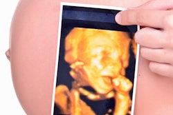
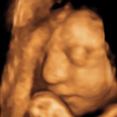
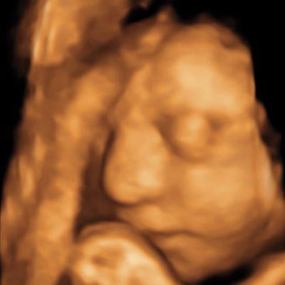 A 3D ultrasound image obtained at 30 weeks in the case of a 34-year-old woman with confirmed Zika virus infection, initially seen for a rash at eight weeks of gestation. Image of the face shows a sloping forehead, compatible with frontal lobe hypoplasia. All images courtesy of Radiology.
While microcephaly is a hallmark of Zika, and most fetuses underwent at least one imaging examination that showed head circumference below the fifth percentile, there are a number of other brain abnormalities that indicate the presence of the disease, according the article. These include abnormalities in ventricular size, gray- and white-matter volume loss, abnormalities in the brainstem, and calcifications.
"It is important for radiologists to understand the type of abnormalities associated with congenital Zika virus infection to aid in recognition of disease and appropriate counseling of patients," the authors wrote.
In all but one of the 17 cases of confirmed Zika infection on prenatal imaging, fetuses had head circumference at or below the fifth percentile on ultrasound performed during the second trimester. Changes in brain parenchyma were also hallmark signs, with a reduction in parenchymal volume that was present on all images.
In addition, 94% of the fetuses in the confirmed Zika group had abnormalities of the corpus callosum, a condition found in 79% of the presumed Zika group. All but one had abnormalities in cortical migration, which means that neurons didn't travel to the proper location in the brain.
What's more, almost all the neonates had intracranial calcifications, most often at the gray-white junction of the brain. And all neonates showed varying abnormalities in cortical development, according to the report.
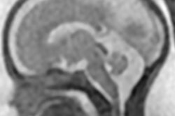
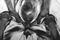
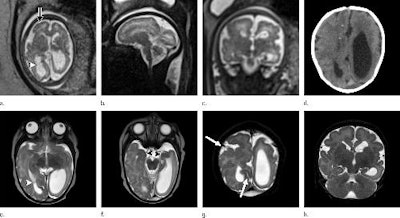 (a) Axial, (b) sagittal, and (c) coronal fetal T2-weighted MR images were obtained in a 29-year-old woman with confirmed Zika virus infection, initially seen for characteristic rash at 12 weeks of gestational age. (d) Axial postnatal CT image and (e-g) axial and (h) coronal T2-weighted MR images were obtained in her neonate. The fetal MR images obtained at 34 weeks (a-c) show asymmetrical ventriculomegaly with a septation in the right occipital horn (arrowhead on a), small frontal lobes, thinning of the occipital parenchyma (left worse than right), underdeveloped sylvian fissures, and regions of thickened cortex, as in the right frontal lobe, which is suggestive of polymicrogyria (arrow on a). There is abnormal, increased signal intensity in the white matter. The postnatal CT image (d) obtained in the 22-day-old neonate shows punctate calcifications at the gray matter-white matter junction and asymmetrical ventriculomegaly. The T2-weighted MR images obtained at 26 days (e-h) show septation in the ventricle (arrowhead on e). Note how the right ventricle has relatively decreased in size compared with the prenatal image, whereas the left ventricle has increased in size. Underrotation of the hippocampus (arrowheads on f) is demonstrated. There is clear asymmetry of the gyral pattern on g, which is relatively smooth in the left occipital region, with abnormal folds in the right occipital and frontoparietal regions (arrows on g). Subependymal cysts are visualized on h, which are not seen on fetal MR images.
"The severity of the cortical malformation and associated tissue changes and the localization of the calcifications at the gray-white matter junction were the most surprising findings in our research," said Dr. Fernanda Tovar-Moll, PhD, in a statement released by RSNA.
Other signs of Zika infection in neonates included a collapsed appearance of the skull, with overlapping sutures and redundant skin folds. These unusual characteristics could be caused by the small brain as it develops, but they also could be due to potential ventriculomegaly leading to a larger head size that then decompresses, and/or brain atrophy that gives the skull a collapsed shape.
Ultrasound can visualize the changes that Zika infection can cause in fetuses, but it may take more than one ultrasound or MRI scan to fully appreciate them, the authors noted.


