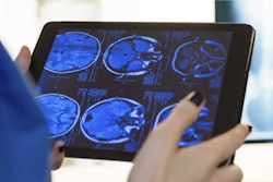
Patients get more out of receiving surgical information via an animated video presentation on an iPad than from a face-to-face consultation with their physician, according to research presented this week at the annual meeting of the European Association of Urology in Munich.
In a randomized controlled trial, a team of researchers from Australia found that 80% of patients favored a video presentation with cartoon animation they could watch on an iPad. Patients may be intimidated and find it difficult to understand the medical language often used by doctors during face-to-face consultations, according to lead researcher Dr. Matthew Winter of Royal North Shore Hospital in Sydney. What's more, busy physicians face time constraints that can affect the quality of consultations or verbal consents for procedures.
"We have found patient's knowledge is greatly improved through the use of portable video media and is their overall preferred method of information delivery compared with standard verbal communication," Winter noted in a statement.
In their study, the Australian team included 88 patients who were facing surgery for acute renal colic. Initially, 45 discussed the surgery with their doctor in a standard face-to-face consultation, while the other 43 received the video presentation with a cartoon animation narrated by a physician. The patients were then asked about their understanding of the medical procedure and their satisfaction with the way the information was presented. Afterward, each group of patients then received the other presentation method.
Of the 88 patients, 71 (80.7%) preferred the video format, while 17 (19.3%) favored the face-to-face meeting. In addition, the video increased patient understanding by 15.5% in comparison with the in-person consultation.
Winter said the researchers are not advocating that portable video media replace the traditional consent process, however.
"Our work shows that there are alternatives to interviews, which can help significantly improve patient understanding, and satisfaction," he pointed out. "Most patients prefer being able to use the portable media devices to a face-to-face consultation, which benefits both clinician and patient through improved quality of care. Portable video media is a useful addition to the informed consent process and I predict will form a crucial component of this process in years to come."


![Examples of ultrasound findings and techniques. (A) Images in a 39-year-old male patient with a mass in the left thigh. The mass is heterogeneous on the B-mode US image (compared with the patient in D) and showed increased microvascularity (superb microvascular imaging [SMI]) and shear-wave elastography (SWE) values. Undifferentiated pleomorphic sarcoma was diagnosed at biopsy (with pleomorphic rhabdomyosarcoma in surgical specimen). (B) Images in an 18-year-old male patient with a mass in the left leg. The mass is hypoechoic on the B-mode image, with no other findings suggestive of malignancy. The lesion is in contact with the cortex of the tibia, which is slightly irregular. CT revealed a doubtful anteromedial tibial erosion. The microvascular study demonstrated high vascularization, suggestive of malignancy. Periosteal Ewing sarcoma was diagnosed with both histologic and immunohistochemical confirmation. (C) Images in a 69-year-old female patient with a lump growing on the outside of the left leg. Multiple SWE examinations were performed (please note the high values obtained in the measurements, whereas the color map highlights the stiffness relative to adjacent tissues). SMI showed areas of increased vascularization to target for sampling. Undifferentiated spindle cell sarcoma was diagnosed at biopsy, with residual leiomyosarcoma in the surgical specimen after neoadjuvant therapy. (D) Images in a 56-year-old female patient with a mass in the right thigh. The mass is heterogeneous at both B-mode ultrasound (similar to patient A) and MRI (coronal T2-weighted spectral attenuated inversion recovery [SPAIR]; T1-weighted pre-contrast and postcontrast imaging), which even shows uptake after the administration of paramagnetic contrast material, which is traditionally suggestive of malignancy. Low values at SMI and elastography are suggestive of benignity. Spindle cell lipoma was diagnosed at biopsy, with atypical spindle cell lipomatous tumor in the surgical specimen.](https://img.auntminnieeurope.com/mindful/smg/workspaces/default/uploads/2026/08/images-radiol250278fig2.APCFLSvX6p.jpg?auto=format%2Ccompress&fit=crop&h=100&q=70&w=100)


![Examples of ultrasound findings and techniques. (A) Images in a 39-year-old male patient with a mass in the left thigh. The mass is heterogeneous on the B-mode US image (compared with the patient in D) and showed increased microvascularity (superb microvascular imaging [SMI]) and shear-wave elastography (SWE) values. Undifferentiated pleomorphic sarcoma was diagnosed at biopsy (with pleomorphic rhabdomyosarcoma in surgical specimen). (B) Images in an 18-year-old male patient with a mass in the left leg. The mass is hypoechoic on the B-mode image, with no other findings suggestive of malignancy. The lesion is in contact with the cortex of the tibia, which is slightly irregular. CT revealed a doubtful anteromedial tibial erosion. The microvascular study demonstrated high vascularization, suggestive of malignancy. Periosteal Ewing sarcoma was diagnosed with both histologic and immunohistochemical confirmation. (C) Images in a 69-year-old female patient with a lump growing on the outside of the left leg. Multiple SWE examinations were performed (please note the high values obtained in the measurements, whereas the color map highlights the stiffness relative to adjacent tissues). SMI showed areas of increased vascularization to target for sampling. Undifferentiated spindle cell sarcoma was diagnosed at biopsy, with residual leiomyosarcoma in the surgical specimen after neoadjuvant therapy. (D) Images in a 56-year-old female patient with a mass in the right thigh. The mass is heterogeneous at both B-mode ultrasound (similar to patient A) and MRI (coronal T2-weighted spectral attenuated inversion recovery [SPAIR]; T1-weighted pre-contrast and postcontrast imaging), which even shows uptake after the administration of paramagnetic contrast material, which is traditionally suggestive of malignancy. Low values at SMI and elastography are suggestive of benignity. Spindle cell lipoma was diagnosed at biopsy, with atypical spindle cell lipomatous tumor in the surgical specimen.](https://img.auntminnieeurope.com/mindful/smg/workspaces/default/uploads/2026/08/images-radiol250278fig2.APCFLSvX6p.jpg?auto=format%2Ccompress&fit=crop&h=112&q=70&w=112)