Diagnosis of skin cancer currently requires a biopsy, which is costly, often causes permanent scarring, and may expose the patient to the risk of infection. New research projects are demonstrating that various optical imaging technologies have the potential to provide noninvasive diagnostic tools for dermatologists.
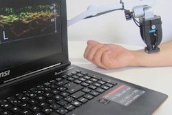
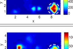
Three-dimensional multispectral optoacoustic mesoscopy is one of these promising technologies. Researchers at the Technical University of Munich have recently used 3D multispectral optoacoustic mesoscopy to reveal melanin and blood oxygenation in the skin on the palm and lower arm of a healthy male volunteer. Their experiment demonstrated the potential ability of the technique to create 3D maps of the skin, providing quantitative readings on oxygenation and lesion penetration.
This imaging technology may help identify biochemical or metabolic alterations of skin due to disease. Changes in oxygen saturation in tumor microenvironments are associated with disease phenotypes including melanoma and cancer progression. Oxygen levels in ischemic wounds are indicators of healing processes. Hypoxic conditions may also identify systemic sclerosis and tissue inflammation (Journal of Biophotonics, January 2016, Vol. 9:1-2, pp. 55-60).
Optoacoustic mesoscopy uses high-frequency ultrasound (20-180 MHz) to obtain high-resolution (3-30 ïm) images of optical absorption to a depth of 1-5 mm, with skin penetration determined by the optical wavelength range and ultrasound frequencies employed.
Optical coherence tomography, which is also being evaluated as a means to identify basal cell carcinoma, can only reach a depth of approximately 1.5 mm (Journal of Clinical Aesthetic Dermatology, October 2015, Vol. 8:10, pp. 14-20). Another unique feature of optoacoustic mesoscopy is its ability to resolve tissue chromophores and photo-absorbing moieties, including melanin, hemoglobin, fluorochromes, and metal nanoparticles, at high resolution.
Skin-specific system
Mathias Schwarz, a PhD student, and colleagues designed a multispectral optoacoustic mesoscopy system specifically for 3D evaluation of human skin. In previous work, this research team showed that raster-scan optoacoustic mesoscopy can visualize different layers in human skin. The team was able to show the epidermal layers, epidermal-dermal junction, and the vascular network of the dermis.
"In our latest study, we bring new insight into optoacoustic mesoscopy of human skin by adding the spectral dimension," Schwarz said. "Thus, multispectral optoacoustic mesoscopy allows us to quantify blood oxygenation in human skin."
The researchers acquired absorption spectra of four different skin compartments over a wavelength range of 460-650 nm. The skin compartments included upper and lower dermal vasculature, epidermis, and the outermost layer of the epidermis (stratum corneum).
The system developed by Schwarz and colleagues was designed to minimize motion of the human body. This was a necessity, they explained, because "on timescales in the order of several seconds, motion is inevitable when imaging human skin in vivo. Quantification of chromophores without precise coregistration of multispectral data will be prone to motion artefacts and is therefore not meaningful."
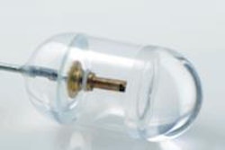
To achieve this, the system uses interleaved multispectral nanosecond laser pulses coupled into a fiber bundle and directed onto the sample. The ultrasound transducer and illumination fibers are raster-scanned in tandem in the x-y plane by two piezo-electric stages. Reflections from every laser pulse are collected by a photodiode, and trigger the data acquisition card. The ultrasound signal is amplified before digitization. A desktop computer controls the entire acquisition protocol and the digitization.
The per-pulse tuneable laser enables coregistration of spectral datasets, from which volumetric multispectral datasets can be used to create 3D absorption maps of melanin, oxyhemoglobin, and deoxyhemoglobin. The researchers were able to calculate the oxygenation level of the vascular network from the absorption maps of both oxy- and deoxyhemoglobin.
"Multispectral optoacoustic mesoscopy brings new sensing ability to dermatology that can impact detection and diagnosis. Multispectral optoacoustic mesoscopy could enable for the first time 3D mapping throughout the skin, allowing information on lesion penetration and quantitative readings on oxygenation," the authors wrote. Schwarz added that "in many skin conditions, especially skin cancer, tissue and blood oxygenation are expected to change."
Schwarz told medicalphysicsweb that the researchers have so far shown the potential of raster-scan optoacoustic mesoscopy and multispectral optoacoustic mesoscopy only in healthy volunteers. They next hope to bring their new technology to clinics to study the imaging capabilities of multispectral optoacoustic mesoscopy in patients with skin cancer and other skin diseases. "If multispectral optoacoustic mesoscopy confirms its diagnostic potential in the clinic, it will certainly be worthwhile to commercialize the technology."
© IOP Publishing Limited. Republished with permission from medicalphysicsweb, a community website covering fundamental research and emerging technologies in medical imaging and radiation therapy.


