A proof-of-concept experiment with a combined high-intensity focused ultrasound (HIFU) and local harmonic motion (LHM) imaging catheter has shown that the minimally invasive catheter can function as a therapeutic and monitoring tool for cardiac ablation. The study, from the University of Toronto, demonstrated the catheter's potential to produce and detect cardiac-ablation lesions.
The current gold standard in cardiac-ablation therapy is radiofrequency (RF) ablation. This RF treatment is limited to the production of transmural lesions in the ventricles, and treatment-monitoring techniques are currently not available for most RF-ablation procedures.
A fully ultrasound-based system has the potential to produce and detect deep lesions, according to lead author Mathew Carias, a graduate student in the Department of Medical Biophysics at the University's Sunnybrook Research Institute. Such lesions are created to destroy diseased tissues, such as abnormal cardiac cells that cause the heart's electrical signal to malfunction and cause irregular heartbeats (Ultrasound in Medicine and Biology, 29 September 2015).
LHM imaging, an elastographic technique that uses both therapeutic and diagnostic ultrasound transducers, is based on the principle that harmonic motion can be estimated and imaged in tissues in which a time-varying force has been externally applied with focused ultrasound transducers. Importantly, it has the ability to continuously monitor lesion formation without discontinuing therapeutic ultrasound.
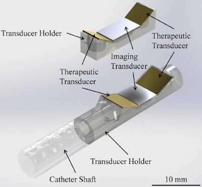 Local harmonic motion (LHM) devices. Top: The first prototype used to determine if a catheter-sized device is capable of producing and detecting displacements. The acrylic holder holds all three transducers. Bottom: LHM 15 French (5 mm-diameter) catheter device in a rapid prototyped transducer holder. Reprinted from Ultrasound Med. Biol., Mathew Carias and Kullervo Hynynen ©2015, with permission from Elsevier.
Its ability to measure lesion formation at multiple tissue depths overcomes current challenges related to knowing lesion-depth penetration during cardiac-ablation therapies. It could also provide a more cost-effective approach to treatment monitoring, compared with techniques such as magnetic resonance thermometry.
For these reasons, Carias and co-author Kullervo Hynynen constructed and tested a combined HIFU and LHM catheter. They initially created a catheter-sized LHM device to detect tissue displacements, testing this on phantoms and ex vivo cardiac samples. They next created a side-facing intravascular catheter in which two embedded angled therapeutic transducers were placed alongside an imaging transducer. This design enabled lesion formation at the same time as acquisition of amplitude motion-mode ultrasound images that enabled tissue displacement to be estimated.
The researchers used this second device to perform an in vivo experiment using a six-month-old 40 kg pig. The left ventricle was used to attempt three lesions on its endocardial surface, and the right ventricle was used to create five lesions on its epicardial surface. Sections of the ventricles that had been exposed to ultrasound were evaluated for overall depth of coagulation into the ventricle wall.
Findings and future plans
The transducers were characterized according to their overall acoustic power output and pressure-field formation, and were determined to have an efficiency of approximately 25%. Measurements in cardiac tissues before and after lesion formation revealed a decrease in displacement amplitude, corresponding to an increase in tissue stiffness. Continuous measurements throughout the sonications showed decreased amplitudes in both ex vivo and in vivo experiments.
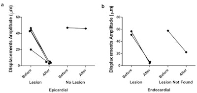 Local harmonic motion amplitudes measured before and after sonication for all epicardial (a) and endocardial (b) in vivo sonications. Reprinted from Ultrasound Med. Biol., Mathew Carias and Kullervo Hynynen ©2015, with permission from Elsevier.
"Four of five epicardial attempts gave rise to lesion formation on the surface, and two of three endocardial attempts gave rise to lesions," the authors wrote. "Endocardial lesions were difficult to locate for histologic examination because of the tortuosity of the intracardial morphology."
Based on their results, the authors recommend that additional experiments are performed to enable clinical use of the device. These include the formation of larger lesions, design of a therapy controller that will enable automatic lesion detection through an online surgical platform, and increased robustness of lesion localization. A controller with automatic amplitude threshold detection would detect when treatment is complete and halt the sonication. They also recommended an investigation to determine the threshold value that gives rise to the most successful treatments.
Carias told medicalphysicsweb that he and his colleagues are currently developing protocols and methods that could be used as a treatment controller, and are also developing threshold values. "I am currently determining what exactly is needed to make a catheter for clinical use. This includes making slight design changes to the catheter itself so that it could be used more safely in a clinical environment," he explained.
© IOP Publishing Limited. Republished with permission from medicalphysicsweb, a community website covering fundamental research and emerging technologies in medical imaging and radiation therapy.
