A previous boss told me once that you never read the really important stuff in textbooks. You learn it by doing the job, she assured me.
This desire to tell you what you don't read in the textbooks and to fill in the gaps is probably what inspired Dr. Paul McCoubrie to write down his own golden rules of radiology. He produced his first set of rules in early 2013, and that article proved very popular. Now he's produced some more, and you can get them here.
Why do so many medical doctors love golf? That's a really tough question to answer, but a group of researchers from Leeds -- a U.K. center of excellence for sports imaging -- has taken a long, hard look at the type of injuries incurred by professional golfers. To read about their findings, go to our MRI Community, or click here.
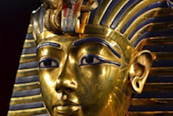
Because of the political situation in Egypt, radiological investigations on the country's ancient mummies had to be suspended more than four years ago, but we asked top radiologist Dr. Ashraf Selim to reflect on how this research is conducted and what's been learned over the years. Visit our CT Community, or click here.
High-quality cost-effectiveness studies from recognized imaging experts remain in short supply in Europe, so new Swiss-led cardiac research looks certain to generate considerable interest. Find out more in our Cardiac Imaging Community, or by clicking here.
Meanwhile, our editorial board member Dr. Anagha Parkar from Norway has provided a case report about a 70-year-old woman with intermittent obstipation and diarrhea. To take the test, click here.
The French congress of radiology, JFR, begins on Friday, so keep a look out for our news coverage of the congress. If you missed our preview of the event, click here.